Fig. 12.1
H-E staining. The number of cells is increased and the collagen bundles are more densely packed with less space in between in the capsule of frozen shoulder. Scale bar = 100 μm

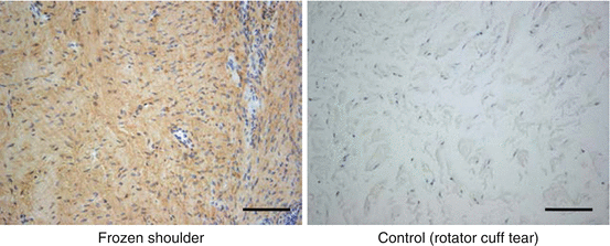
Fig. 12.2
Immunohistochemistry. Strong staining of type I collagen is observed in the capsule of frozen shoulder. Scale bar = 100 μm
Cytokines and growth factors related to fibrosis and inflammation increased in the joint capsule from frozen shoulder. Transforming growth factor beta (TGF-β), platelet-derived growth factor (PDGF), hepatocyte growth factor (HGF), interleukin-1-beta (IL1-β), IL-6, tumor necrosis factor-alpha (TNF-α), vimentin, secreted protein, acidic, cysteine-rich (SPARC, osteonectin), cyclooxygenase (COX) 1, COX-2, and intercellular adhesion molecule-1 (ICAM-1, CD54) increased in frozen shoulder [6, 8, 11, 14, 15, 20]. No up-regulation of matrix metalloproteinasis-1 (MMP-1), MMP-2, MMP-3, MMP-9, and MMP-14 was reported [5, 6]. Tissue inhibitor of metalloproteinase-1 (TIMP-1) increased but not for TIMP-2 and TIMP-3 in frozen shoulder [6]. Besides increased vascularity [6, 8, 24], an angiogenic factor of cystein-rich, angiogenic inducer, 61 (CYR61) increased in the joint capsule from frozen shoulder [6]. A decrease of blood supply from anterior humeral circumflex artery may affect an increase of angiogenic factors and hypervascularity related to a poor posture, frequently seen in patients with frozen shoulder [7]. Neurogenesis [24] and increased expression of neuronal proteins, such as substance P and calcitonin gene-related peptide (CGRP), were observed in frozen shoulder [6].
Same trends in the joint capsule were observed in synovial tissues. Related to fibrosis and inflammation, TNF-α, IL1-α, IL-1β, IL-6, TGF-β, PDGF, fibroblast growth factor (FGF), COX-2, and MMP-3 increased in the synovial tissues from frozen shoulder [10, 15, 20]. For angiogenesis, vascular endothelial growth factor (VEGF) increased in frozen shoulder [9, 11, 21].
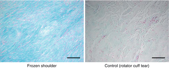
Besides fibrosis and inflammation, chondrogenesis is one of the main pathologies of frozen shoulder [6, 18]. Although alcian blue staining intensity increased, there were no chondrocyte-like cells in frozen shoulder (Fig. 12.3). Gene expressions of aggrecan (ACAN), type II collagen (COL2A1), and type X collagen (COL10A1) increased in frozen shoulder [6]. From DNA microarray analysis, some genes related to fibrosis such as collagen type I (COL1A1) and collagen type XIII (COL13A1), chondrogenesis such as FBJ murine osteosarcoma viral oncogen homolog (FOS), ACAN, FOSB, and COL10A1, angiogenesis such as CYR61 were up-regulated in frozen shoulder [6].


Fig. 12.3
Alcian blue staining. Proteoglycan, a major component of cartilage, is richly observed in the capsule of frozen shoulder (blue staining portion). Scale bar = 100 μm
12.2 Laboratory Tests
There is no specific blood test for frozen shoulder. The serum triglyceride and cholesterol levels were significantly elevated in frozen shoulder [4]. For type 2 diabetic patients, the blood sugar, both fasting and 2 h after breakfast, HbA1c and serum triglyceride levels significantly increased in patients with frozen shoulder compared to those without it [22]. Serum IgA and lymphocyte transformation to phytohemagglutinin significantly reduced [2]. Serum levels of ICAM-1 [14], TIMP-1, TIMP-2, and TGF-β1 were significantly higher, but MMP-1 and MMP-2 levels were significantly lower in frozen shoulder [16]. After intensive stretching, serum levels of MMP-1 and MMP-2 increased but TIMP-1 and TIMP-2 decreased compared with supervised neglect in frozen shoulder [16].
References
1.
2.
Bulgen D, Hazleman B, Ward M, McCallum M. Immunological studies in frozen shoulder. Ann Rheum Dis. 1978;37(2):135–8.CrossRefPubMedCentralPubMed
Stay updated, free articles. Join our Telegram channel

Full access? Get Clinical Tree


