Fig. 10.1
(a) Zero-degree abduction of glenohumeral–capsular complex. (b) The capsule showing high stresses (light blue color) in various positions of representative patient with frozen shoulder
10.6 Altering Scapular Position Associated with Frozen Shoulder
Our study was also linked to evaluate the positional change of scapula and humerus with respect to the rib cage during two different shoulder abduction angles. A better understanding of positional differences of the scapula and humerus during glenohumeral movement is needed to better define the functional change of periscapula muscles and kinematic alteration of scapulothoracic force couple in patients with various degrees of frozen shoulder.
Thus, we compared the positional difference between scapulae obtained at maximum abduction position and 0° abduction in three patients with typical frozen shoulder. The averaged scapula–humeral angle of the affected shoulder and contralateral shoulder were 34 ± 11° and 92 ± 7°, respectively. The abductions of the scapula of the affected shoulder and contralateral shoulder were 58 ± 7° and 38 ± 8°, respectively. The posterior tipping of the affected shoulder was 25 ± 5° (Fig. 10.2).
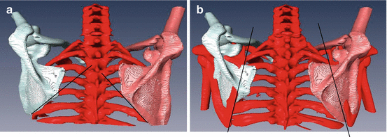
Fig. 10.2
A 3D abduction image of representative case in frozen shoulder (a) The left scapula (affected side) showing an increase in upward rotation. (b) Overlap image between two different positions indicating a decrease in medical rotation of scapula at abduction
Therefore, the decrease of posterior tipping known generally in case of frozen shoulder should be verified again through an in-depth study. Also, the medial rotation of the scapula in the affected shoulder was decreased compared with the contralateral scapula in the same degree of glenohumeral angle. However, it is not obvious whether the cause of decreased medial rotation is due to the imbalance of scapular thoracic muscular activity or scapular stiffness itself. The scapula stiffness is another important factor that interferes with the natural shoulder kinematics in frozen shoulder. This concept was first suggested by Hamada et al. What they found in patient with frozen shoulder with passive mobilization test was a decreased range of motion in medial rotation, posterior tilt, and downward rotation. They considered scapular stiffness and muscular tightness as major factors rather than capsular contracture in frozen shoulder.
10.7 Summary and Conclusion
Pathomechanical cascade in frozen shoulder has yet to be fully understood. Although a few studies have pointed out abnormal scapular location according to glenohumeral abduction using electromagnetic tracking device in frozen shoulder patient, that method has an innate limitation in evaluating detailed motions except upward and downward rotation [24]. Likewise, EMG study focusing on periscapular muscle activity did not provide an integral insight into changing the way of activity in both scapulohumeral and scapulothoracic muscle groups [10]. Thus, abnormal biomechanics in frozen shoulder needs to be further investigated.
According to our result and speculation based on our 3D kinematics, early abutment of humeral head against acromion which in turn causes failed obligatory external rotation was speculated to be an initiator of pathologic cascade in pathogenic mechanics of frozen shoulder. The change in position of humeral head causes another change in the axis of rotation of both the humerus and scapula, resulting in unpredictable periscapular muscular action.
In conclusion, pathomechanics of frozen shoulder characterized by glenohumeral joint limitation has a complicated pathogenesis made with the contracture of glenohumeral capsule, the periscapular stiffness, and the imbalance of strength of muscles surrounding the scapula.
References
1.
Basmajian JV, Bazant FJ. Factors preventing downward dislocation of the adducted shoulder joint. J Bone Joint Surg Am. 1959;41:1182–6.PubMed
Related posts:

Stay updated, free articles. Join our Telegram channel

Full access? Get Clinical Tree


