Patellar Tendon Repair
Albert M. Tsai
INDICATIONS/CONTRAINDICATIONS
Isolated rupture of the patellar tendon is a rare injury occurring primarily in patients <40 years old. Most ruptures occur as the result of a rapid forceful contraction of the quadriceps muscle with a flexed knee, and a history of stumbling or giving way of the knee can often be elicited along with a sudden pop and acute onset of pain. It has been demonstrated that normal tendons usually do not rupture without significant trauma (4). A certain degree of tendon degeneration is often present, the result of cumulative microtrauma or the degenerative changes associated with aging. The injury can also be associated with systemic diseases such as chronic renal failure, rheumatoid arthritis, systemic lupus erythematosus, or diabetes mellitus and occurs with little or no trauma in these patients. Rarely, iatrogenic patellar tendon rupture has been reported after harvesting the middle third of the tendon for anterior cruciate ligament reconstruction (1,6).
Surgical repair of an acutely ruptured patellar tendon is the standard of care and is necessary to reestablish continuity of the extensor mechanism of the knee. Neglected injuries can lead to proximal retraction of the quadriceps and patella with resultant adhesions and extensor mechanism insufficiency. Contraindications to acute surgical repair include acute infection, medical comorbidities that make the surgical risk prohibitive, and soft tissue loss that makes primary repair impossible.
PREOPERATIVE PLANNING
The first step in assessing the injured knee is a thorough history and physical examination. There is generally swelling and ecchymosis about the knee as well as tenderness along the patellar tendon or at the inferior aspect of the patella. There may also be a palpable defect in the tendon. Occasionally, the patient will be able to perform a straight leg raise maneuver through an intact extensor retinaculum. However, the diagnosis should still be apparent as there will usually be an extensor lag and weakness when compared to the uninjured extremity.
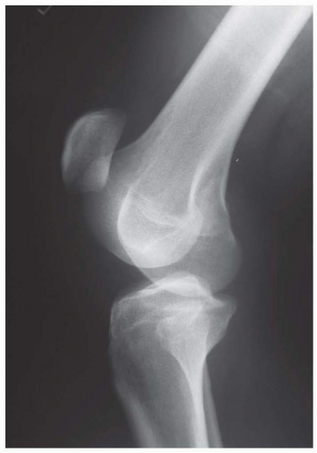
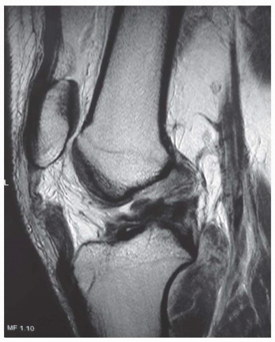
Imaging of the knee should include routine roentgenograms to rule out fractures. On a lateral view of the knee, patella alta can be seen (Fig. 19.1). Magnetic resonance imaging (MRI) is not usually necessary in acute injuries but may be helpful in chronic neglected ruptures, in those few acute cases where the diagnosis is unclear, or to diagnose suspected concomitant intra-articular pathology (Fig. 19.2).
SURGERY
Patient Positioning
The patient is placed in the supine position, and a small bump is placed under the ipsilateral hip to keep the patella pointing toward the ceiling. A tourniquet is placed on the proximal thigh but is usually not inflated during the procedure. The entire extremity is then sterilely prepped and draped free.
Technique
A midline skin incision is made centered over the defect in the patellar tendon. If the disruption is an avulsion off the inferior pole of the patella, the incision may extend more proximally; in the case of a midsubstance tear or tibial tubercle avulsion, the incision may extend more distally. Routine dissection is carried sharply down
through skin and subcutaneous tissue. At this point, the knee joint is frequently visualized through the ruptured tendon. The joint is thoroughly irrigated to remove old hematoma and soft tissue debris, and the articular surfaces can be examined for any injury. If possible, the paratenon should be identified and incised in order to further dissect out the patellar tendon in preparation for repair.
through skin and subcutaneous tissue. At this point, the knee joint is frequently visualized through the ruptured tendon. The joint is thoroughly irrigated to remove old hematoma and soft tissue debris, and the articular surfaces can be examined for any injury. If possible, the paratenon should be identified and incised in order to further dissect out the patellar tendon in preparation for repair.
Most commonly, the patellar tendon is avulsed from the inferior aspect of the patella, along with tears of the medial and lateral retinaculum (Fig. 19.3). The frayed, tattered stump of patellar tendon is sharply débrided with dissecting scissors or a sharp scalpel in order to freshen up the tendon and remove any nonviable tissue. Next a Rongeur is used to débride the inferior pole of the patella, removing soft tissue remnants and providing a healthy bleeding bone surface for the repaired tendon. A total of two 2 FiberWire sutures are woven into the end of the tendon, using a Krackow-type locking stitch. One suture is woven into the medial half of the tendon, working from proximal to distal and then back up in the medial central portion of the tendon. The second suture is similarly woven down the lateral half of the tendon and back up the lateral central portion of the tendon, leaving four suture strands exiting the tendon (Fig. 19.4). These sutures are marked with hemostats so that they can
later be passed through drill holes in the patella. For ease of identification, the central sutures can be cut shorter than the peripheral ones and clamped together (Fig. 19.5).
later be passed through drill holes in the patella. For ease of identification, the central sutures can be cut shorter than the peripheral ones and clamped together (Fig. 19.5).
Stay updated, free articles. Join our Telegram channel

Full access? Get Clinical Tree


