Fig. 19.1
Classic “slapped” cheek rash of erythema infectiosum (fifth disease) on face and torso (Reprinted from Schneider AP, Naides SJ. Human Parvovirus Infection. Am J Family Physician. 39:165, 1989. With permission from American Academy of Family Physicians)
Differential Diagnosis
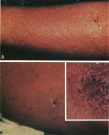
Bright-red “slapped” cheeks are highly suggestive of B19 infection. Macular or maculopapular rash is nonspecific and may be seen in a number of viral infections and drug eruptions. A history of exposure to B19 via a family contact or during a known outbreak is a helpful clue. Individuals with occupations that allow exposure to small children, e.g., school teachers or pediatric nurses, are at risk. Outbreaks typically occur in the winter and early spring, but outbreaks at other times of the year occur as do sporadic cases. A history of sudden-onset, symmetric, small joint polyarthralgia or polyarthritis should suggest the possibility of B19 infection. Rubella infection can present in the exact same manner with similar rash and polyarthritis in a college age teen or young adult but is less common than B19 infection. Failure to confirm a recent B19 infection by the presence of serum anti-B19 IgM antibody should prompt consideration of testing for serum anti-rubella IgM antibody. Purpura may resemble many causes of purpura due to coagulopathy. Petechiae may resemble many causes of petechiae due to thrombocytopenia. Henoch-Schönlein purpura is suggested by the localization of purpura to the abdomen, buttocks, and lower extremities in the setting of arthritis, gastrointestinal symptoms, and/or hematuria following an antecedent respiratory illness; multiple triggers are known including B19 infection [1–4]. The differential diagnosis of the vesiculopustular eruption includes herpes simplex, varicella zoster, and vaccinia virus infection. (Smallpox is also in the differential but not a routine consideration given its eradication from world populations.) “Gloves and socks” acral erythema is seen in B19.
Biopsy
Histological changes are not considered diagnostic. The putative receptor for B19, globoside, is present in the plasma membrane of endothelial cells. B19 may induce apoptosis in target cells nonpermissive for B19 replication through expression of its nonstructural protein, NS1, a helicase capable of damaging host cell genomic DNA. Electron microscopy may show B19 in lesional endothelial cells [6]. Catastrophic skin vascular endothelial cell injury has been described in B19 infection in which in situ reverse transcription and hybridization showed B19 RNA transcripts [3]. Vesiculopustular lesions show ballooning degeneration of keratinocytes with a mixed inflammatory cell infiltrate that may contain numerous eosinophils. Local edema, dilated vessels, and erythrocyte extravasation suggest local capillary leak. A lymphohistiocytic infiltrate with markedly abnormal atypical cells showing enlarged, hyperchromic nuclei may be present (Figs. 19.2 and 19.3) [7].


