Chapter 12 Paediatric Proximal Ulnar Fractures
Introduction
Fractures of the proximal ulna comprise several types of olecranon fractures together with fractures involving the coronoid process. The reported incidence is between 4% and 7% of paediatric elbow fractures1–3 and they are often seen as part of combined more complex elbow injuries. Their incidence may vary greatly, however, even between different regions of the same country. For example in the Malmö area of Sweden they represent only 1.7% of paediatric elbow fractures, which is equivalent to an annual incidence of 0.8 olecranon fractures per 10 000 children.2
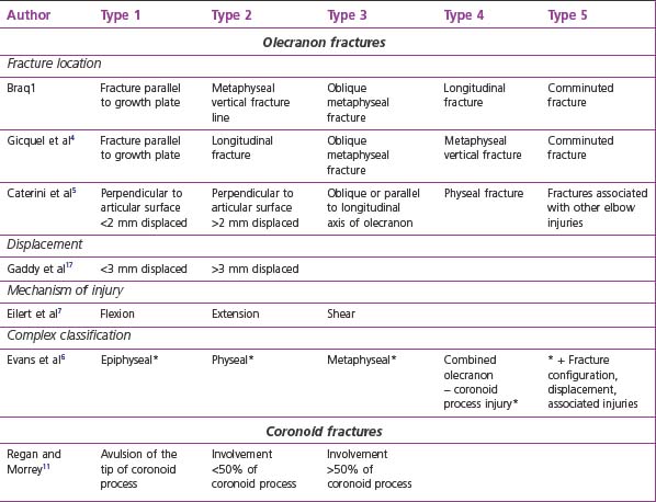
There are no widely accepted classification systems for olecranon fractures (Table 12.1). Bracq1 described a five-type classification system which was modified by Gicquel et al4 and which is based on the direction and localization of the fracture line. Another five-type classification system was introduced by Caterini et al.5 Their five categories differentiated between the direction of the fracture line, the degree of fracture displacement and the presence of associated injuries. A much more complex classification is described by Evans and Graham.6 They put emphasis on the anatomical site, the fracture configuration, the degree of intra-articular displacement and the type of associated injuries. The principal classification concentrates on four anatomically defined types: epiphyseal, physeal, metaphyseal and combined olecranon–coronoid process injuries. A simple two-group classification was advised by Papavasiliou et al.3 They separated intra-articular from extra-articular fractures, dividing the intra-articular fractures into four subgroups according to the degree of fracture displacement.
The mechanism of injury that produces different reproducible fracture patterns has also been used as a functional classification.7 In flexion type injuries the rare avulsion fracture and the much more frequent metaphyseal fractures of the olecranon occur. Fractures that result from injuries with an extended elbow usually result in greenstick type fractures of the olecranon with longitudinal fracture lines. They have associated injuries in 40–70% of cases. I am a firm believer in the functional classification and finds it very practical and useful.
Summary Box 12.1
Proximal ulna fractures account for 4–7% of paediatric elbow fractures.
They comprise olecranon and coronoid process fractures.
Several classification systems exist (Table 1), of which the functional classification based on the mechanism of injury is practical and helpful in planning treatment.
Background/aetiology
Olecranon fractures can occur after a fall onto the flexed or extended elbow.
In fractures that result from a flexion type injury a tension force acutely pulls on the posterior part of the olecranon mainly through the power of the triceps brachialis muscle but with assistance from the biceps, which produces a flexion force. This leads to a separation of the olecranon by a fracture line perpendicular to the longitudinal axis of the ulna, either through the metaphyseal area of the proximal ulna or through the proximal ulnar growth plate. The fracture line usually extends into the joint, making the injury an articular fracture. A rare flexion type injury that results from a direct blow to the olecranon creates shear forces at the area of impact, leading to a vertical fracture line with an intact proximal radio-ulnar joint. In these fractures the posterior periosteum does not disrupt and has the effect of an internal tension band.7
The extension type injury usually needs an additional valgus or varus stress to cause an olecranon fracture. In a fall on the outstretched hand the elbow joint is overextended with the olecranon locked in the olecranon fossa.1 Without additional valgus or varus stresses this injury mechanism most likely results in a supracondylar fracture. With additional valgus or varus stresses the olecranon may fracture in a greenstick type manner, producing longitudinal fracture lines that may extend well into the proximal ulnar shaft region. This fracture pattern is triggered by the specific anatomical properties of the growing proximal ulna. The olecranon represents a metaphyseal bony region with a strong periosteal sleeve. The periosteum may prevent true fracture displacement, allowing only for some bending in either a valgus or varus direction. This type of injury pattern makes associated lesions very likely.
Presentation, investigation, treatment options
Flexion type injury
Clinical presentation
The clinical presentation of a flexion type olecranon injury is characterized by local tenderness and swelling associated with an inability to actively extend the elbow. In children this may be difficult to test adequately because of pain and swelling. However, a multicentre prospective study has shown a high sensitivity of the elbow extension test at detecting any type of elbow fracture. In children there was an almost 50% likelihood of having sustained a fracture if the child was unable to actively extend the elbow.8 In those cases with major displacement of the fragments the gap between the bony pieces may be palpated.
Treatment options
The degree of fracture displacement will determine the treatment modality. Many of these fractures are not displaced or are mildly so, requiring plaster immobilization alone, or closed reduction followed by plaster immobilization with the arm in 80–90° of flexion.1 Some authors recommend plaster immobilization in elbow extension, but we have not found this to be either an appropriate or superior method of immobilization.
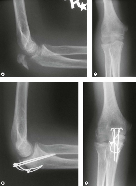
Moderately displaced fractures or unstable mildly displaced fractures that redisplace after closed reduction are best managed by closed reduction and percutaneous pinning by K-wires. Postoperatively an above-elbow cast in neutral is applied for 4 weeks. Severely displaced or irreducible fractures do need open reduction and internal fixation in order to achieve an anatomical position. The fixation method of choice in this situation is the classic K-wire fixation with AO tension band wire (Fig. 12.1).9


