Total Elbow Arthroplasty

Chapter 43 Total Elbow Arthroplasty


Indications and implant types currently available


The clinical indication for elbow arthroplasty is pain that is uncontrolled by simple analgesia, non-steroidal antiinflammatory drugs or other modalities. Pain at rest with associated sleep disturbance is a particularly good reason for surgery. Added to this is loss of movement and strength, with consequent diminution in function and the ability to work.


Of the various conditions affecting the elbow the most common reason for total elbow arthroplasty is inflammatory arthritis (rheumatoid/psoriatic). Other indications include elbow trauma in the elderly, particularly comminuted unreconstructable intra-articular distal humeral fractures, distal humeral non-unions and posttraumatic arthritis. In addition total elbow arthroplasties may occasionally be performed for osteoarthritis and tumour reconstruction.


Contraindications are generally relative and, if anything, are diminishing due to improvements in surgical technique and implant design. Active infection remains an absolute contraindication, but previous infection is not a contraindication provided that the infection has been satisfactorily eradicated. The complete absence of elbow flexion is also a contraindication in so much that an elbow prosthesis will only be of value if it is inserted into an elbow that is capable of active movement. Poor skin quality is a relative contraindication and should be addressed prior to surgery by appropriate flap cover. An unstable or severely deformed elbow is a contraindication for an unlinked prosthesis and will require a linked implant due to the high risk of instability postoperatively. Patients who undergo total elbow arthroplasty must be prepared to accept a permanent restriction on lifting and work activities with heavy manual labour remaining an absolute contraindication for this type of surgery.


Implants currently available can be classified into three types: linked, unlinked and modular (prostheses that can be linked or unlinked depending on circumstances). The most widely used arthroplasty in America is the Coonrad–Morrey, which is a linked implant, while in Europe and the rest of the world, linked and unlinked implants are used equally. The more recent modular designs which can be inserted as unlinked or linked implants are an attempt to increase the options available.


Linked arthroplasties rely on the sloppy hinge for stability with a pin and polyethylene construct through which movement occurs. The inbuilt ‘sloppiness’ equates to an average of 8° varus/valgus laxity at the articulation. While having many advantages, a particular problem with this type of device is that when the elbow functions at the extremes of varus or valgus, forces are transmitted to the linkage mechanism, resulting in polyethylene wear. If this continues or is extreme then these forces are transmitted directly to the stem and cement bone interface. As a consequence the humeral or ulnar component will loosen. Alternatively the bushings wear with disassembly of the implant. In contrast the unlinked total elbow arthroplasty has no intrinsic link and relies on the soft tissues, particularly the collateral ligaments, for its integrity. Generally these implants mimic to a greater or lesser degree the normal articular surfaces of the humerus and ulna. Again the extremes of varus and valgus can result in increased polyethylene wear. In general, however, it is felt that these implants lessen the stresses on the bone cement interface allowing the capsular ligaments to transfer forces and therefore decrease the risk of mechanical loosening.


When considering elbow joint design it is important to understand constraint, especially as unlinked implants are often referred to as unconstrained devices. The Oxford English Dictionary defines constraint as ‘limitations imposed on motion’. Whilst soft tissue structures, particularly the collateral ligaments, contribute significantly to stability around the normal elbow, there is some inbuilt incongruity of the articular surfaces that allows 6–8° of varus valgus. Any implant design that does not have this inbuilt incongruity is more constrained even if it is unlinked. Indeed such an implant can be more constrained than a linked implant with a sloppy hinge. As might be expected, there is significant variation between implants.1


Due to the high failure rate of the original surface replacements, modern implants have a stem on both the humeral and ulna components. In addition, most components are cemented in place. Humeral fixation can be augmented in a number of ways of which an anterior flange is perhaps the most popular. It is thought that this produces better force transfer across the humerus, although recently this has been disputed by Quenneville et al.2 Others3,4 have used a wedge design for the humeral component, the edges of which sit in both supracondylar ridges and resist rotation forces. In addition implant fixation has been improved by the use of better cementing techniques which include cement restrictors (bone blocks or manufactured devices), bone preparation and pressurized cement insertion.


With regard to ulnar components, all implants must accommodate the normal valgus angulation of the ulnar stem relative to the olecranon fossa. This makes the provision of separate implants for left and right sides essential.5 For most unlinked implants the ulnar component is the site of the high-density polyethylene. This may be cemented directly into bone as in the Souter–Strathclyde prosthesis, although more recent designs frequently incorporate a metal backing.


Implant stem length varies in different prostheses and may affect long-term survival of the arthroplasty. Using the Souter–Strathclyde prosthesis, Trail et al,6 showed that when the 7 cm humeral stem was compared with the 3.5 cm stem, the longer stem had a lower incidence of loosening.


The incorporation of a radial head replacement as part of a total elbow arthroplasty remains controversial. Some of the early designs had a radial head prosthesis, but these were soon withdrawn due to problems with loosening. The obvious mechanical advantage of sharing the forces across the elbow has been off-set until recently by the design difficulties. One of the latest implants, however, is now a three-piece arthroplasty comprising humeral, ulnar and radial head components (Latitude-Tournier). Whether this will stand the test of time or indeed confer any long-term advantage on the arthroplasty remains to be seen.


Finally, the long-term outcome of any elbow arthroplasty depends not only on the implant itself but also on the skill of the surgeon. The effect of intraoperative placement and alignment of the components was analysed by Shah et al7 who found that for the Souter–Strathclyde prosthesis correct alignment and positioning of the humeral implant was crucial to its long-term survival. Indeed there was little latitude for malalignment. While this is essential for unlinked implants to avoid the risk of dislocation, it is obviously important that all implants are inserted in a satisfactory and reproducible manner. To this end, many of the newer designs have a full set of instrumentation allowing bone resection to be undertaken in a standard fashion.


Surgical techniques and rehabilitation


A number of surgical approaches for elbow arthroplasty have been described in Chapter 6. The technique utilized, however, generally depends on surgeon preference and particularly training, as well as the type of implant being inserted. The author’s personal preference is a technique that has evolved at Wrightington Hospital over the last 20 years. This is based on a posterior approach, which involves taking down a tongue of triceps.


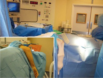
The operation is performed in a laminar airflow theatre under general anaesthesia and with an inter-scalene block. The patient is placed in the lateral decubitus position with the arm suspended over a gutter (Fig. 43.1). A high tourniquet is applied allowing access to the elbow joint and distal humerus. The patient’s position should not impede the surgeon placing the arm in full extension and flexion during the surgical procedure. Prophylactic antibiotics are administered prior to inflation of the tourniquet in order to reduce the risk of infection.


A dorsal longitudinal incision is made down to and including the deep fascia. Skin flaps are raised and reflected in a radial and ulnar direction. By using a longitudinal incision and raising thick flaps the incidence of skin edge necrosis has virtually been eliminated. The only location the author would avoid placing the incision is directly over the tip of the olecranon.


Flaps are dissected out to both the medial and lateral epicondyles. On the medial side the ulnar nerve is identified and superficially decompressed within the cubital tunnel. In primary surgery I no longer routinely undertake an anterior transposition since I have found that leaving the ulnar nerve in its normal bed has reduced the incidence of postoperative ulnar nerve problems. An anterior transposition is, however, undertaken if there is bony deformity or prominence and when revision surgery is performed.


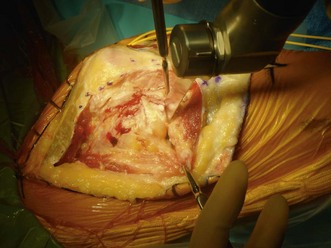
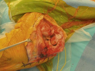
With the triceps exposed, a longitudinal incision is made just to the radial side of the midline again avoiding the tip of the olecranon. The incision is carried distally to expose the radial head (Fig. 43.2), which is routinely excised proximal to the annular ligament. The triceps and its attachment to the olecranon and distal ulna are then reflected as one piece. This is facilitated by using a sharp osteotome, particularly over the tip of the olecranon. The whole flap is then reflected medially exposing the fat pad and dorsal capsule of the elbow joint. Whilst the fat pad is retained, the capsule is excised. The tip of the olecranon is also removed. With further release of the posterior parts of the medial and lateral collateral ligaments, the elbow is dislocated (Fig. 43.3). This is further aided by undertaking a coronoid osteotomy and, if appropriate, releasing the anterior capsule. At the end of this part of the procedure, the only structures intact are the anterior parts of both collateral ligaments. The importance of these structures has been demonstrated in a number of studies.8,9


The implant is then inserted as per the manufacturer’s instructions. For an unlinked arthroplasty alignment of the components is crucial, not only in the anteroposterior and latero-medial planes, but also in depth of insertion of the implant. Accurate depth positioning will maintain stability yet still allow a full range of motion. The principle is that with the trial implants in situ there should be total stability with a full range of motion. If this is not the case the surgeon must re-evaluate the trial implant position in order to correct the situation. The same principles apply for a linked implant, although more of the collateral ligaments can be sacrificed in order to gain accurate positioning. A malaligned linked prosthesis will result in increased polyethylene wear and early loosening/disarticulation.


Once the arthroplasty has been inserted the triceps tendon must be reconstructed with transosseous sutures. The advantage of this approach is that triceps continuity and length is maintained helping stability but also allowing early mobilization. The deep fascia is then repaired and the skin sutured. I always leave two drains in situ in order to prevent haematoma formation and reduce the risk of infection.


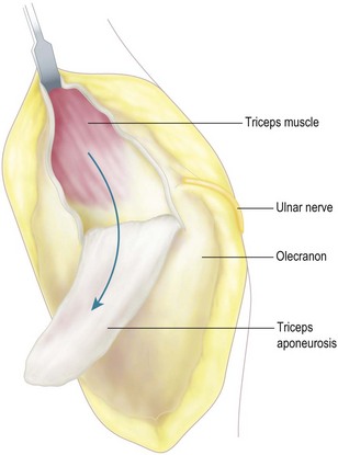
The original surgical approach that I used involved the same skin incision but gained access to the elbow joint by taking down a distally based flap of the triceps tendon (Fig. 43.4). Distally the incisions were extended to release the posterior parts of the medial and lateral collateral ligaments. The dorsal capsule was then incised and the elbow joint exposed. Whilst this approach was used successfully for many years, it undoubtedly resulted in some ongoing weakness of the triceps tendon which is why I no longer use it routinely. The only indication now would be if there was a significant loss of flexion preoperatively and at the end of the procedure lengthening of the triceps tendon was required.


An alternative lateral approach is favoured by some surgeons.10 This is based on a modified Kocher approach. The operation is performed under general anaesthesia with the patient in the lateral position. A tourniquet is used and the patient given prophylactic intravenous antibiotics. Ulnar nerve decompression, if necessary, is undertaken through a separate medial incision.


The skin incision is made between the lateral epicondyle and the olecranon and utilizes any previous incisions to avoid skin necrosis. The lateral collateral ligament is detached from its humeral origin and the anterior and posterior capsules released. This allows the elbow to be opened as a hinge and the medial collateral ligament preserved. When the implant has been inserted, the lateral collateral ligament is reattached with sutures through bone. It is obviously important that during this process the correct tension of the collateral ligament is maintained so that the joint is stable with a full range of motion.


Ljung et al11 reported a high incidence of ulnar nerve palsy with this approach but there were few incidences of instability.


Rehabilitation


At the end of the operation the elbow is immobilized in a dorsally fitting splint the position of which is somewhat controversial. Some surgeons immobilize the elbow at 90° while others favour more extension. The rationale for the latter being that extension is more difficult to regain after total elbow replacement than flexion. Added to this, and perhaps more importantly, many unlinked elbow replacements, at least in our experience, are more stable in extension rather than flexion.


The day after surgery the splint is removed and a wound inspection and dressing change undertaken. If drainage is minimal, the drains are removed. Early mobilization is then instigated providing the surgeon feels that the arthroplasty is stable. This involves flexion and extension together with pronation and supination exercises. These should be performed four to five times a day both passively and actively. It is important that during this time the collateral ligaments, particularly the medial ligament, are protected thus reducing the risk of instability. The shoulder and wrist should also be kept mobile.


Between exercise periods and particularly at night in bed the elbow should be protected in a splint. This splint may have to be remoulded on several occasions as the swelling subsides. Generally it is worn for a minimum of 6 weeks. If during this period the patient has difficulty regaining extension, the splint can be placed on the volar side of the elbow stretching the arm into further extension. Three weeks after surgery light function can commence out of the splint using the arm to feed and dress. It is, however, important that no loaded activities are undertaken and that the patient continues to use the splint in crowds, when travelling and at night. At 6 weeks limited strengthening exercises can be commenced. A life time ban is recommended, however, on lifting more than 2.5 kg (5 lb). Added to this, joint protection techniques for activities of daily living are discussed and emphasized.

Stay updated, free articles. Join our Telegram channel

Sep 8, 2016 | Posted by in MUSCULOSKELETAL MEDICINE | Comments Off on Total Elbow Arthroplasty

Full access? Get Clinical Tree

Get Clinical Tree app for offline access