Otorhinolaryngology

Chapter 19 Otorhinolaryngology


Emergencies


Key Points


Epiglottitis


Epiglottitis (or “supraglottitis”) is a condition that requires prompt attention by the physician. Epiglottitis results from bacterial (and rarely viral) infection of the supraglottic structures, that is, the epiglottis and arytenoid cartilages. A high level of suspicion is necessary to make a diagnosis and avoid significant morbidity. Rapid decompensation and complete loss of the airway are the sequelae of most concern. The physician should always be suspicious when a patient presents with fever, sore throat, and difficulty swallowing, and when the severity of oropharyngeal physical findings is not in proportion to the symptoms. Croup, tonsillitis, peritonsillar abscess, and other neck infection may be incorrectly diagnosed in these patients. Epiglottitis occurs mainly in children age 2 to 7 years, although infants, older children, and adults can be affected. Mortality rates of 6% to 7% have been reported in adults.


Signs and symptoms of epiglottitis include rapidly developing sore throat, high fever, restlessness, and lethargy. A “supraglottic,” muffled voice is common. Many patients have difficulty with their saliva and drool. Classically, these patients are in a sitting position leaning forward, because this position tends to alleviate obstructive symptoms from the supraglottic swelling. They may show signs of “air hunger” or may have stridor.


Differential diagnosis includes tonsillitis, peritonsillar abscess, retropharyngeal abscess, airway foreign body, and croup. Physical examination with laryngoscopy is extremely useful in differentiating these diagnoses. Endoscopy should not be performed if there is concern of impending airway obstruction. Endoscopy will typically show erythema and edema of the epiglottis and arytenoid cartilages. Other findings include laryngeal tenderness on neck palpation, although palpation should be avoided when the diagnosis is being considered.


Any time the diagnosis of epiglottitis is in question, otorhinolaryngologic (ear-nose-throat, ENT) and infectious disease consultations are warranted. Placement of a tongue depressor has been known to precipitate acute airway obstruction and should be avoided entirely if epiglottitis is strongly suspected. Differentiation from croup can be difficult because there is considerable overlap of symptoms (Table 19-1) (Berry and Yemen, 1994). A lateral extended neck radiograph can help in the diagnosis. X-ray evidence includes the classic “thumbprint” sign. If epiglottitis is suspected or lateral neck radiography is confirmatory, the patient should be taken to the operating room (OR) for orotracheal intubation in the presence of an anesthesiologist and an otorhinolaryngologist. In any case of airway obstruction, cricothyrotomy or tracheotomy can be lifesaving, because orotracheal intubation can be difficult and sometimes impossible. Some patients, usually adults, may be treated expectantly with intravenous (IV) medications and intensive care unit (ICU) observation as long as personnel are available for control of the airway if necessary. If airway stability is questionable, observation is not recommended.


Table 19-1 Distinguishing Features of Epiglottitis and Croup


Feature Epiglottitis Croup
Cause Bacterial Viral
Age 1 year to adult 1 to 5 years
Location of obstruction Supraglottic Subglottic
Onset Sudden (hours) Gradual (days)
Fever High Low grade
Dysphagia Marked None
Drooling Present Minimal
Posture Sitting Recumbent
Toxemia Mild to severe Mild
Cough Usually none Barking, brassy, spontaneous
Voice Clear to muffled Hoarse
Respiratory rate Normal to rapid Rapid
Larynx palpation Tender Not tender
Clinical course Shorter Longer

From Berry FA, Yemen TA. Pediatric airway in health and disease. Pediatr Clin North Am 1994;41:153.


After control of the airway is achieved, cultures of the epiglottis should be obtained and directed antibiotics instituted. Haemophilus influenzae type b (Hib) is common and can be beta-lactamase producing. Other, less common organisms include beta-hemolytic streptococci, Streptococcus pneumoniae, and Staphylococcus aureus. Antibiotics should be administered parenterally; effective antibiotics include cefotaxime, ceftriaxone, ampicillin plus sulbactam, or ampicillin plus chloramphenicol. Steroids can be useful for edema and inflammation, but their effectiveness has not been proved in controlled studies.


The incidence of epiglottitis in children is decreasing since the introduction of the Hib vaccine in the late 1980s. However, the incidence has remained stable or slightly increased in adults.


Peritonsillar Abscess


A peritonsillar abscess is the accumulation of pus in the peritonsillar space that surrounds the tonsil. The same organisms responsible for common tonsillar infections— Streptococcus and Staphylococcus species and anaerobes—are also found in peritonsillar abscesses.


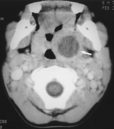
The typical signs and symptoms of peritonsillar abscess include fever, sore throat for 3 to 5 days, dysphagia, odynophagia, and a muffled, “hot potato” voice. Trismus is extremely common. Examination confirms asymmetric tonsils and peritonsillar edema and erythema. The soft palate and uvula are swollen and displaced away from the side of the abscess. It is often difficult to distinguish between abscess and peritonsillar cellulitis. If possible, it is helpful to palpate because fluctuance indicates a loculation of pus. Diagnosis is often made by clinical impression, but computed tomography (CT) can be confirmatory and useful when the diagnosis is uncertain (Fig. 19-1).


If untreated, a peritonsillar abscess may spontaneously drain, progress to involve the deep neck, or even lead to airway obstruction. The most important part of the treatment is drainage of the abscess cavity by needle aspiration, incision and drainage, or tonsillectomy. Cultures of the aspirate can be obtained, and broad-spectrum antibiotics should be started. Appropriate antibiotics include ampicillin-sulbactam (Unasyn) or clindamycin (Cleocin). Many patients present with dehydration, and parenteral fluids should be given if necessary. Analgesics should be prescribed as needed. One or two doses of IV corticosteroids may be given to decrease inflammation and pain.


Children presenting with peritonsillar abscess should be admitted to the hospital. Treatment with IV hydration and parenteral antibiotics is appropriate initially. Patients with peritonsillar cellulitis/phlegmon or early abscess often demonstrate a rapid response to treatment, whereas those with a well-formed peritonsillar abscess do not improve. Drainage is necessary in nonresponders. Abscesses in cooperative adults can be drained under local anesthesia in the emergency department (ED) or office and treated in an outpatient setting. Children usually require general anesthesia for drainage, and a tonsillectomy may also be performed. An elective tonsillectomy is often recommended for any patient with a peritonsillar abscess to prevent recurrence, especially with a history of recurrent tonsillitis, although few, if any, controlled studies support this recommendation.


Foreign Bodies


Swallowing or aspiration of objects is most common in children but also occurs in the adult population. These objects can become lodged anywhere in the upper aerodigestive tract.


Esophagus


The most common location for esophageal foreign bodies is at the level of the cricopharyngeal muscle. Other regions include the anatomic narrowings of the esophagus, such as the gastroesophageal junction, and the area of indentation of the esophagus by the left main stem bronchus and the arch of the aorta. Coins are by far the most common objects found in the esophagus in children. Chicken or fish bones are more common in adults.


The diagnosis of an esophageal foreign body is primarily based on the medical history and physical examination, with the aid of radiologic studies. Parents might witness ingestion of the foreign body and subsequent coughing, gagging, refusal to eat, or drooling. Often, however, the incident goes unwitnessed, and reliance on other diagnostic techniques is necessary.


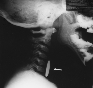
Plain radiographs (including lateral films) are often diagnostic in pediatric patients because most esophageal foreign objects are radiopaque (Fig. 19-2). Other radiologic findings that can suggest a foreign body include increased soft tissue density in the prevertebral space, mediastinal widening, air-fluid levels in the esophagus, and paraesophageal air. Disk batteries require a high index of suspicion because they can cause significant tissue injury and lead to esophageal perforation if they are not removed emergently. They have a classic appearance when viewed laterally, approximating a dime resting on a nickel (similar to the appearance of Fig. 19-2).


If there is sufficient evidence of an esophageal foreign body, ENT consultation is indicated for rigid esophagoscopy and removal. When radiolucent objects have been ingested, contrast esophagography might be indicated, although esophagograms can give a false-negative result and can also complicate visualization during rigid esophagoscopy.


Airway


As with esophageal foreign bodies, airway foreign bodies are much more common in infants and young children. Many deaths from foreign-body aspiration occur in the home before medical intervention can be administered. The most frequently aspirated foreign bodies are foods, with nuts leading the list. Foreign bodies aspirated into the airway are usually found lodged in the bronchial tree but can also be found in the larynx or trachea. If the event is witnessed and results in complete airway obstruction, a Heimlich maneuver should be administered; however, the event is often not witnessed. Symptoms can include hoarseness, persistent cough, wheezing, or stridor if the foreign body is lodged in the trachea or larynx. Because the potential for morbidity and mortality is substantial, this condition requires urgent diagnosis and timely intervention to prevent catastrophe.


Any time a small child presents with wheezing or noisy breathing without a previous history of reactive airway disease, an airway foreign body should be included in the differential diagnosis. Typically, patients and parents recount a transient episode of coughing during eating that then subsided; the patient might even be symptom free for a time, then later have symptoms such as coughing or wheezing.


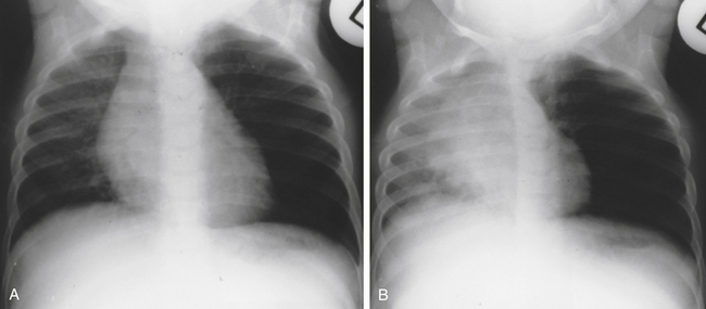
The most important diagnostic step for identifying a foreign body is a high index of suspicion. Careful auscultation of the lung fields is essential because subtle asymmetric differences may be found. Because most airway foreign bodies are radiolucent, chest radiographs can be normal, but abnormalities such as hyperinflation, atelectasis, or pneumonia can be present (Fig. 19-3). If plain radiographs are equivocal or normal and the patient is in stable condition, airway fluoroscopy can be helpful.


Consultation with a physician experienced in foreign body removal is required. Definitive treatment of airway foreign bodies is direct laryngoscopy and rigid bronchoscopy to indentify and remove the object.


Epistaxis


Although epistaxis is usually nothing more than a minor annoyance, some episodes are severe enough to require urgent medical attention and intervention. In rare cases, epistaxis can also be a life-threatening emergency.


Predisposing factors for epistaxis include trauma (facial trauma or self-inflicted digital trauma), dry weather, hypertension, bleeding dyscrasias (factor deficiencies, hereditary hemorrhagic telangiectasia, lymphoproliferative disorders), anticoagulation therapy (acetylsalicylic acid, heparin, warfarin), and intranasal tumors. Special consideration should be given to adolescent boys with recurrent epistaxis and nasal obstruction, because these symptoms might be the result of a juvenile nasopharyngeal angiofibroma. These are benign but locally aggressive tumors. All these potential risk factors should be considered because they must be addressed to treat the patient appropriately.


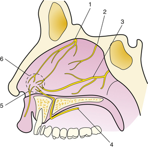
Epistaxis is classified according to its location. Bleeding from the anterior nasal cavity is most common and usually originates from a rich plexus of vessels at the anterior septum called Kiesselbach’s plexus (Fig. 19-4). Bleeding from this location, although troublesome, is less likely to be severe and is usually easier to control than posterior epistaxis. Posterior epistaxis originates from the posterior two thirds of the nasal cavity and can be quite severe and much more difficult to control.


Initial management of epistaxis includes assessment and stabilization of vital signs. Rarely, severe bleeding can lead to airway compromise or hemodynamic compromise, or both, especially in patients with underlying cardiopulmonary dysfunction. The airway should be assessed and stabilized, urgently if necessary. Hypertension, if severe, should be controlled, with care taken to avoid subsequent hypotension. Hematologic studies, including complete blood count (CBC), prothrombin time, and partial thromboplastin time, should be ordered. Intravenous access should be established, allowing administration of fluids as well as IV medications, if necessary during treatment.


Treatment


Effective treatment requires adequate visualization and patient cooperation. The level of intervention by the primary care physician depends on level of experience, comfort level, and availability of appropriate supplies and equipment. ENT consultation should be considered when any of these prerequisites cannot be met. The patient should be reassured and given an explanation of the planned treatment. This results in better cooperation and decreases patient anxiety, improving treatment success. A bright headlight, nasal speculum, large nasal (Frazier tip) suction, and bayonet forceps are required. If the patient is monitored and stable, a small dose of IV narcotics titrated for analgesia and anxiolytic effect may be given. Extreme caution should be taken not to oversedate the patient. Also, instrumentation of the nose can lead to a significant vasovagal response that may be accentuated in a hypovolemic patient given narcotics. It is advised to err on the side of caution with regard to narcotic medication both during and after treatment.


All clots should be suctioned. They can be quite tenacious and require forceps for removal. The nasal cavity should next be topically anesthetized and decongested (a mixture of 4% lidocaine and phenylephrine works well). If a bleeding site is easily identified, it may be cauterized with a silver nitrate stick.


Anterior Packing


If suction, decongestion, and cautery do not stop the bleeding and the site is still thought to be anterior, an anterior nasal pack should be placed. This can be done with 0.5-inch petroleum gauze coated in antibiotic ointment. Alternatively, a variety of preformed packs are available. Merocel nasal packs or prepackaged inflatable packs are usually readily available and quite effective. Remembering that the nasal cavity extends posteriorly from the nostril and not superiorly facilitates placement. The pack should be coated in antibiotic ointment before placement.


Placement of the pack can be quite uncomfortable for the patient. Discomfort can be minimized by ensuring optimal decongestion and topical anesthesia of the nasal cavity. If using a Merocel pack, it is sometimes helpful to hydrate and expand the pack before placing it. This can be done with sterile saline or phenylephrine. Although the pack appears quite large after it is expanded, it decompresses readily and slides in easily once it is covered in antibiotic ointment. The entire length of the pack needs to be grasped with the bayonet forceps if this technique is employed. Preexpansion of the pack also minimizes further abrasions and bleeding that can occur with placement of the firm pack. This technique is especially advantageous if a septal deviation or spur exists on the bleeding side. Regardless of what type of pack is used, care must be taken not to distort or overstretch the nasal ala (nostril) once the pack is in place. This causes significant discomfort and can result in necrosis of the nostril.


After the pack is in place, the patient should be observed for further bleeding. If the patient remains stable, has no significant comorbid conditions, and has only a unilateral anterior pack, the patient may be discharged with mild narcotic pain medications and antibiotics for prophylaxis against toxic shock syndrome and sinusitis. The pack should be removed 2 to 5 days later, with instructions to use nasal saline and nasal ointment liberally for the next 2 weeks. Recurrent bleeding should prompt an ENT consultation.


Posterior Packing


If the bleeding is not controlled with an anterior pack, the origin of the bleeding may be posterior, requiring an anterior-posterior pack. An anterior-posterior pack is inserted similar to a nasogastric tube, then inflated with the minimum amount of saline to stop the bleeding. The recommended maximum amount of inflation should not be surpassed.


If a preformed anterior-posterior pack is not available or not effective, a traditional anterior-posterior pack may be placed. The posterior pack is necessary essentially to seal off the posterior nasal cavity and provide a buttress to prevent the anterior pack from slipping posteriorly. A Foley catheter and balloon is slid into the nasal cavity and serves as the posterior pack and buttress. Once the Foley catheter is in place, it is inflated with 5 to 10 mL of saline (or air) and pulled snugly to seal the posterior nasal choana. A large anterior pack is then placed, which should quickly control the bleeding.


Placement of a posterior pack requires experience, and even if it is successfully done, ENT consultation is indicated to assist with further bleeding, pack removal, and to monitor for pack complications. Anterior-posterior packs are associated with significant patient discomfort and potential complications during and after insertion. Local complications, including necrosis of the alae, septum, and palate, can occur, and close observation is required to prevent them. Hypoxemia can also result. Supplemental oxygen and monitoring of oxygen saturation is indicated. Patients with preexisting cardiopulmonary disease require closer observation, often in the ICU. The patient might require judicious doses of narcotics for pain and antibiotics for infection prophylaxis.


Head and Neck Trauma and Respiratory Embarrassment


Key Points


The ABCs of trauma should be remembered when treating a patient with cervicofacial trauma. This includes evaluation and treatment of airway, breathing, circulation, and cervical spine. The most pressing issue after significant face, head, or neck trauma is the potential for respiratory compromise, secondary to several causes. Altered mental status can lead to aspiration of blood or secretions with or without central hypoventilation. Comminuted facial fractures (midface or mandibular) can distort the oral and pharyngeal airway sufficiently to cause obstruction. An undetected expanding pharyngeal or neck hematoma can cause airway obstruction by extrinsic compression of the trachea or pharynx. Blunt or penetrating neck injuries can cause laryngeal fracture, bleeding, or hematoma, leading to critical airway obstruction.


Potential airway obstruction must be addressed quickly because complete obstruction can progress rapidly. The diagnosis is clinical because hypoxemia and carbon dioxide retention are late signs. Extensive facial edema or ecchymosis should arouse concern for facial fracture. A muffled voice can be the result of expanding hematoma. Laryngeal or tracheal injury should be suspected if the patient has a change in the voice, hemoptysis, subcutaneous emphysema, or stridor.


Stabilization of a compromised airway should be accomplished as soon as possible. Endotracheal intubation may be attempted, with plans for emergent cricothyrotomy as necessary. If time permits, the on-call anesthesiologist, trauma surgeon, or otorhinolaryngologist should be consulted to assist in airway management. Blind intubation (especially nasotracheal) or insertion of a laryngeal mask airway (LMA) is not recommended because this further compromises the already tenuous airway if intubation is unsuccessful. Although tracheotomy is the preferred procedure when endotracheal intubation is impossible or contraindicated, cricothyrotomy is also acceptable and can be lifesaving.


There is potential for significant blood loss after severe head and neck trauma. Intravenous access should be established and volume replacement initiated quickly. Bleeding from facial wounds can be controlled with direct pressure and suture ligation of arterial bleeding. Management of epistaxis is discussed earlier. Bleeding from the neck, or evidence of expanding hematoma, implies a major vessel injury and requires immediate operative exploration by a trauma surgeon, vascular surgeon, or otorhinolaryngologist.


Unrecognized pharyngeal and esophageal injury can result in life-threatening infection. These injuries might not be obvious on initial evaluation and require a very high index of suspicion. Contrast studies and endoscopy are usually required to confirm the diagnosis. Treatment can include repair of the injury or external drainage to allow healing.


Isolated facial injuries are rarely life threatening but still can result in significant bleeding and, rarely, airway compromise and permanent disability. Significant facial trauma should be evaluated in the ED. The potential for intracranial and cervical spine injuries should be considered when major facial injuries are present. Trauma of the periorbital region requires ophthalmologic evaluation. All lacerations should be inspected, cleaned, and sutured. Antibiotics should be used if contamination is likely.


Deeper injuries can result in facial nerve transection. If facial nerve weakness is detected, plastic surgery or ENT consultation is necessary for expedient nerve exploration and repair. The parotid salivary duct can also be injured and requires repair over a stent. Facial fractures should be evaluated with CT (both axial and coronal images). Possible mandibular fractures should be evaluated with plain x-ray films, including panographic (Panorex) films. Overlooked and untreated facial fractures can result in significant long-term functional and cosmetic deficits. Oral surgery consultation is sometimes required, especially with injury to the teeth or altered dental occlusion.


The Ear


Key Points


Otalgia


While the vast majority of patients with otalgia have an otologic cause, the clinician must recognize that otalgia may be referred. Sensory innervation of the ear includes cranial nerves V, VII, IX, and X, and therefore disorders of structures with similar innervation can cause otalgia. It is imperative that the physician not simply attribute otalgia to an ear infection unless the physical examination supports this diagnosis. Otalgia can result from dysfunction of the nose, sinuses, oral cavity, pharynx, larynx, dentition, temporomandibular joints, and salivary glands. These structures must be thoroughly assessed, especially if the examination of the ear appears normal. This is especially true in smokers, whose initial symptom of laryngopharyngeal carcinoma may be otalgia. Otolaryngologic referral for laryngoscopy may be indicated with suspected referred otalgia. (See eBox 19-1 and eBox 19-2 online at www.expertconsult.com for differential diagnosis of otalgia.)


Vertigo


Key Points


The sense of balance or equilibrium occurs when there is normal and harmonious function of several systems and organs in the body. These include the musculoskeletal system, the cardiovascular system, the central nervous system, the eyes, and the ears. Abnormal function of any of these can result in the sensation of dizziness or disequilibrium. The term vertigo is reserved to describe a perceived sensation of motion, usually spinning, of the person relative to the environment, or vice versa. Causes of disequilibrium can be categorized into one of three groups: peripheral (inner ear or labyrinthine), central nervous system (CNS), or systemic (e.g., cardiovascular, metabolic). Although not pathognomonic of a labyrinthine disorder, true vertigo most often indicates aberrant function of the inner ear.


Because patients use “dizzy” to describe many sensations, the actual sensation is best clarified by a detailed history (Box 19-1). The major studies on the causes of persistent dizziness, from Drachman and Hart (1972) to Davis (1994), all describe four diagnostic categories: lightheadedness, presyncope, disequilibrium, and vertigo. The investigators all conclude that the most common cause of persistent dizziness is a peripheral vestibular disorder (38%-56% of cases) followed closely by a psychogenic disorder (6%-33%). In about 25% of patients, the complaint is the result of the combined effects of multiple sensory deficits, medications, or orthostasis, leading to complaints of presyncope, lightheadedness, or disequilibrium. Finally, central vestibular etiologies are unusual and represent less than 10% of all causes.


A thorough medical history allows the physician to distinguish between true vertigo (a sensation of spinning) and other sensations, such as presyncope, lightheadedness, and unsteadiness. The physical examination and laboratory evaluation are guided by the accuracy of the history. A sensation of vertigo originates from within the vestibular system but can be either peripheral (vestibular nerve and inner ear) or central (cerebellum, brainstem, thalamus, and cortex).


Questions regarding hearing and neurologic deficits can help elicit which part of the vestibular system is involved (Wiet et al., 1999) (see eTable 19-1 online). Peripheral vertigo tends to be episodic, whereas central vertigo is constant. Neurologic symptoms or loss of consciousness do not occur with peripheral vertigo but are possible with central vertigo. Nystagmus, which is labeled by the direction of the fast component, can be present in both types of vertigo and can be horizontal or rotary; vertical nystagmus occurs only in central vertigo.


The physical examination should include assessment of orthostatic blood pressure changes, a complete ocular examination, tuning fork tests (Weber’s and Rinne’s), pneumatic otoscopy (elicits vertigo in patients with perilymphatic fistula), balance tests (Romberg’s), gait (including tandem walking), and cranial nerve evaluation. The Dix-Hallpike maneuver (see eFig. 19-3 online) is especially helpful in diagnosing benign paroxysmal positional vertigo (BPPV). Head movement always worsens the feeling of true vertigo. If it does not, the dizziness can be attributed to a cause other than vestibular dysfunction.


Laboratory testing can include an audiogram if no specific cause of vertigo can be found after the medical history and physical examination. Electronystagmography (ENG) is an objective study of the vestibular system and can help localize a vestibular lesion. Electrodes placed about the eye sense the movements of nystagmus as either spontaneous or initiated by maneuvers such as caloric testing, positioning, optokinetics, and pendulum tracing. A brain MRI scan is indicated in patients with unilateral otologic symptoms and in those unresponsive to treatment. Blood tests, when necessary, can include CBC, rapid plasma reagin (RPR), vitamin B12 level, folate level, drug screens, and heavy metal testing when indicated.


Meniere’s Disease


Meniere’s disease is characterized by episodic severe vertigo lasting hours, with associated symptoms of unilateral roaring tinnitus, fluctuating low-frequency hearing loss, and aural fullness. Typical onset is in the fifth decade of life. The cause is uncertain but is speculated to result from allergic, infectious, or autoimmune injury. The histopathologic finding includes endolymphatic hydrops, which is thought to be caused by either overproduction or underresorption of endolymph in the inner ear.


Meniere’s disease is a clinical diagnosis mostly based on history. Testing may be obtained to support the diagnosis and rule out other disorders. Audiometry often demonstrates a low-frequency sensorineural hearing loss. An FTA-ABS test may be obtained to rule out syphilis. ENG may demonstrate a unilateral peripheral vestibular weakness on caloric testing. When the diagnosis is uncertain, a brain MRI with contrast is obtained to evaluate for a retrocochlear lesion. The differential diagnosis of Meniere’s disease includes acute labyrinthitis, neurosyphilis, labyrinthine fistula, autoimmune inner ear disease, vestibular neuronitis, and migraine-associated vertigo.


Although Meniere’s disease has a highly variable clinical course, most patients have long symptom-free periods between clusters of episodes. The majority of patients have an excellent prognosis, with symptoms burning out over several years. However, some patients have a disabling course with frequent and severe attacks. On average, a moderate sensorineural hearing loss is the end result. The disease may become bilateral in about 45% of cases (wide variability exists).


Treatment of an acute episode involves vestibular suppressants and antiemetics. As with any vestibular disorder, vestibular suppressants should be limited for use during acute symptoms because of their addictive potential and impairment of central compensation. Maintenance therapy includes reduction of sodium intake to less than 1500 mg/day and a diuretic such as hydrochlorothiazide-triamterene (Dyazide). Patients are also instructed to minimize caffeine, alcohol, nicotine, and chocolate. Allergy treatment may be helpful in some patients. Most patients have adequate control of symptoms with this regimen.


Patients who fail conservative measures may be candidates for procedures and surgical treatment. Gentamicin, a vestibulotoxic aminoglycoside antibiotic, may be injected transtympanically into the middle ear to permeate into the inner ear. Control of vertigo may result in 90%, but with a risk of hearing loss. Endolymphatic sac decompression or shunting through a mastoidectomy appears to benefit most patients with minimal risk to hearing. Although a generally accepted procedure, adequate studies are lacking on its effectiveness. More invasive interventions, including vestibular nerve section and labyrinthectomy, are reserved for patients with severe disease who do not respond to other measures (Sajjadi and Paparella, 2008).


Benign Paroxysmal Positional Vertigo


The most common cause of peripheral vestibular vertigo in adults is benign paroxysmal positional vertigo. BPPV occurs in all age groups but more often between ages 50 and 70. The incidence of BPPV is 11 to 64 per 100,000 persons per year and is twice as common in women as men (Froehling et al., 1991). It is caused when otoconia particles from the utricle or saccule lodge in the posterior semicircular canal and is also referred to as canalithiasis. This causes the canal to be a gravity-sensing organ, and head movement results in displacement of the otoconia and a sensation of vertigo.


The Dix-Hallpike maneuver reproduces this vertigo in the patient, resulting in nystagmus (see eFig. 19-3). Characteristics of the nystagmus of BPPV include fatigability, a latency period of 1 to 5 seconds before nystagmus begins after the head is moved, short duration of nystagmus from 5 to 30 seconds, and reversal of the nystagmus components when the patient is returned to the sitting position. If these characteristics are not present and treatment is not successful, BPPV cannot be diagnosed. In such a case, a CNS lesion is possible. BPPV can be the residual effect of Meniere’s disease, ear surgery, vestibular neuronitis, or ischemia of the inner ear. Head trauma, even when it is minor, can lead to BPPV. However, one third of cases are idiopathic.


Treatment of BPPV consists of performing repositioning maneuvers with the goal of returning the otoconia to the utricle or saccule (Yacovino, 2009). In addition, the patient may attempt to reposition the otoconia at home by sitting upright on the bed and rapidly lying supine with the affected ear facing downward. After 1 minute, the head should be repositioned with the opposite ear facing downward, and the patient should wait another minute. The patient should then return slowly to the upright seated position and repeat this exercise four more times. The entire process is completed twice daily until the symptoms have abated.


An expert panel convened by the American Academy of Otolaryngology–Head and Neck Surgery Foundation recommended against “routinely treating BPPV with vestibular suppressant medication such as antihistamines or benzodiazepines” (Bhattacharyya et al., 2008). Because of the variability of symptoms, the clinician must judge each case independently. Resolution occurs in a few weeks or months, and the condition is benign, although it can recur.


Labyrinthitis


As with vestibular neuronitis, labyrinthitis causes sudden and severe vertigo. In contrast to vestibular neuronitis, the patient also has tinnitus and hearing loss. The hearing loss is sensorineural, is often severe, and can be permanent. Labyrinthitis is caused by inflammation within the inner ear. The cause is most often a viral infection but can be bacterial. Bacterial labyrinthitis usually results from extension of a bacterial otitis media into the inner ear. A noninfectious serous labyrinthitis can also occur after an episode of acute otitis media. Other, less common causes include treponemal infections (syphilis) and rickettsial infection (Lyme disease).


Symptomatic treatment of labyrinthitis is similar to that for vestibular neuronitis. Antibiotics are recommended if a bacterial cause is suspected. As with acute otitis media, bacterial labyrinthitis can, in rare cases, lead to meningitis. Few other conditions cause the constellation of hearing loss, tinnitus, and vertigo, but cerebrovascular ischemia, meningitis, brain abscess, and encephalitis should all be considered. Although the vertigo should resolve over days to weeks, hearing loss and tinnitus can persist.


Drugs known to be ototoxic can cause acute onset of hearing loss and disequilibrium, although this is not true labyrinthitis. These drugs include salicylates, aminoglycosides, loop diuretics, and various chemotherapeutic agents. This cause should be considered in patients who complain of hearing loss or dizziness while taking these medications.


Tinnitus


Tinnitus is a term used to describe an internal noise perceived by the patient. It is usually, but not always, indicative of an otologic problem. Tinnitus is most often subjective, that is, heard only by the patient. However, it can be objective and heard by the patient and the examiner. In most cases, tinnitus is secondary to bilateral sensorineural hearing loss and requires no further evaluation. In rare cases, tinnitus can be a symptom of a vascular abnormality (aneurysm or arteriovenous malformation), hypermetabolic state, or intracranial mass that, if not evaluated, could result in delayed treatment. Middle ear and rarely external ear pathology can also cause tinnitus, as can numerous medications (Box 19-2). The patient’s medications should be reviewed.


Evaluation of tinnitus begins with a complete medical history, including duration of symptoms, possible inciting event (e.g., acoustic trauma), and accompanying symptoms (e.g., vertigo, hearing loss, headache, vision changes). Specific questions regarding the tinnitus are critical: Is it unilateral or bilateral? What is the quality of the tinnitus (pitch, volume)? Does it sound like a heartbeat or rushing blood? Does it change? A complete ENT evaluation should be performed, and audiometry is mandatory.


In general, if the tinnitus is bilateral, not particularly intrusive, not pulsatile, and associated with symmetric hearing loss, it is likely secondary to the hearing loss itself. The hearing loss requires further evaluation with magnetic resonance imaging (MRI) with contrast if it is asymmetric.


In cases of pulsatile tinnitus with normal otoscopy, magnetic resonance angiography (MRA) is performed to evaluate for vascular abnormalities. If otoscopy identifies a retrotympanic mass, a temporal bone CT is obtained to evaluate for a vascular mass or abnormality. Blood tests can be performed to rule out anemia or hyperthyroidism, which can result in a hypermetabolic state and cause tinnitus secondary to increased blood flow near the cochlea. Auscultation of the neck, periauricular area, and chest may identify a bruit or murmur, indicating a need for a carotid duplex ultrasound study or echocardiogram, respectively. Most cases of arterial pulsatile tinnitus are secondary to atherosclerotic carotid artery disease. Venous pulsatile tinnitus often improves with digital pressure over the internal jugular vein. Etiologies include idiopathic venous hum, a high-riding jugular bulb, or benign intracranial hypertension.


Effective treatment of tinnitus is difficult and usually requires various approaches. Finding and eliminating potential causes (especially pharmaceutical) is imperative. Patients should be counseled to avoid caffeine and nicotine. No single medicine has been proved effective in treating tinnitus. Antidepressants have shown promise, especially if depression coexists. Intravenous lidocaine eliminates tinnitus in some patients but is not practical and has obvious potential side effects. Various homeopathic treatments and nutritional supplements are effective in some cases, but most have not been evaluated in controlled studies. Hearing aids are beneficial in masking the tinnitus if hearing loss exists. Tinnitus maskers can be purchased that essentially drown out the tinnitus with various distracting noises. Biofeedback and a technique called tinnitus retraining therapy are helpful for some patients. These techniques can be learned through various publications or at a tinnitus treatment center. All patients with obtrusive tinnitus are encouraged to join the American Tinnitus Association, the largest tinnitus support group and an excellent source of reliable information.


Disorders of the External Ear


Otitis Externa


The most common cause of pain in the external ear is acute otitis externa. It affects 3% to 10% of the patient population. The pain is caused by inflammation and edema of the ear canal skin, which is normally adherent to the bone and cartilage of the auditory canal. The inflammatory reaction can be caused by bacteria, fungi, or contact dermatitis (see eTable 19-2 online).


Cerumen protects the canal by forming an acidic coat that helps prevent infection. Factors that predispose to otitis externa include absence of cerumen, often from excessive cleaning by the patient; water, which macerates the skin of the auditory canal and raises the pH; and trauma to the skin of the auditory canal from foreign bodies or use of cotton swabs.


When a bacterial organism is suspected, treatment consists of cleaning the ear canal of any debris or drainage and then instilling antibiotic drops with or without steroids. Because the most common bacterial organisms in this infection are Pseudomonas aeruginosa and Staphylococcus aureus, drops containing ciprofloxacin or neomycin/polymyxin B are effective against these pathogens, combined with a steroid to decrease inflammation, pain, and pruritus (Ciprodex, Cortisporin, Coly-Mycin, Pediotic). A recent study found Ciprodex to be more effective against P. aeruginosa than neomycin/polymyxin B/hydrocortisone (Dohar et al., 2009).


The clinician must use judgment in assessing the severity of the infection and treat accordingly. If the infection spreads beyond the auditory canal, oral antimicrobials are indicated. If clinical improvement is not apparent after 48 hours, the patient needs to be reexamined for additional treatment or referral to an otorhinolaryngologist.


Fungal infections compose less than 10% of external otitis cases. The most common fungi are Aspergillus niger and Candida species and are more prevalent in tropical climates. Itching is a more common complaint than pain in fungal ear infections. Thorough cleaning of the ear canal is the primary duty of the physician in this infection. Drops that are effective include 2% acetic acid with or without a steroid. Clotrimazole drops or powder can also be used to treat fungal infections of the canal (van Bolen et al., 2003).


Approximately 90% of necrotizing (malignant) otitis externa is seen in immunocompromised patients such as diabetic patients, patients with acquired immunodeficiency syndrome (AIDS), and those receiving chemotherapy. Systemic antibiotics are mandatory in these cases. Antipseudomonal antimicrobials should be administered intravenously in the hospital setting, and surgical debridement is often necessary. Complications from necrotizing otitis externa include facial nerve palsy, mastoiditis, meningitis, and even death (Quick, 1999).


Other conditions that affect the external auditory canal include impacted cerumen, seborrheic dermatitis, psoriasis, contact dermatitis, and staphylococcal furunculosis. Symptoms and signs include pruritus, edema, scaling, crusting, oozing, and fissuring of the external auditory canal. Treatment of the underlying disease is the primary goal. Corticosteroid preparations are indicated for seborrheic dermatitis, psoriasis, and contact dermatitis. Oral antibiotics and sometimes incision and drainage are required for staphylococcal furunculosis.


External Auditory Canal Foreign Bodies


A common problem seen in family physicians’ offices is a patient with an external auditory canal foreign body. A wide variety of objects can be found. In one study of 191 patients with aural foreign bodies, 27 different objects were discovered (Ansley and Cunningham, 1998). The most common were beads, plastic toys, pebbles, insects (especially cockroaches), popcorn kernels, earrings, paper, peas, cotton, pencil erasers, and seeds. When a patient presents with a chronic dry cough that has not responded to the usual measures, the physician should look for an aural foreign body (causing irritation of the ninth cranial nerve).


Removal of an external auditory canal foreign body is simplified if the object is in the lateral one third of the external auditory canal. Objects within the medial two-thirds pose a greater challenge. A variety of instruments can be used, depending on the object, including cerumen loops, alligator forceps, and otologic-tip suctions. Irrigation with body-temperature sterile water often dislodges the object. Hygroscopic objects such as vegetables, beans, and other food matter can swell and make the object even more impacted and should not be irrigated. Disk batteries should be removed immediately because of the possibility of liquefaction necrosis of the external auditory canal. Aural irrigation is contraindicated because wetting of the battery leads to leakage of electrolyte solution.


Smooth, round objects pose a difficult problem because, in trying to remove them, they are often pushed farther into the canal. Aural irrigation or even cyanoacrylate glue on the tip of a straightened paper clip is effective in removing objects that are difficult to retrieve. Methods to remove cockroaches or other insects include microscope immersion oil, mineral oil, or lidocaine. The effect of mineral oil or microscope immersion oil is to drown the insect, whereas lidocaine tends to make cockroaches crawl rapidly out of the canal (Bressler and Shelton, 1993). Otomicroscopy is often required for safe removal.


Depending on their age, fewer than 35% of patients should require anesthesia. The younger the patient, the more likely anesthesia will be required. Objects with sharp edges are best removed with an operating microscope with the patient under general anesthesia. Complications of foreign body removal include canal wall trauma and tympanic membrane perforation. Immobilization of the patient is the key to successful removal of aural foreign bodies, and at least two assistants are necessary.


Cerumen


Glandular secretions from the outer one third of the external auditory canal and desquamated epithelium combine to form cerumen. Cerumen is necessary to provide a hydrophobic and acidic environment to protect the underlying external ear canal epithelium and prevent infection. The external auditory canal is self-cleaning, with cerumen slowly pushed laterally to the external meatus.


Cerumen impaction is the symptomatic accumulation of cerumen in the external canal or an accumulation that prevents a needed assessment of the ear. Complete occlusion is not necessary. Symptoms may include hearing loss, tinnitus, pruritus, fullness, otalgia, cough, odor, and dizziness. Impaction often results from instrumentation with cotton-tipped applicators, which should be discouraged. Elderly patients with changes to external canal epithelium, patients with external canal abnormalities (e.g., osteomas, exostoses, stenosis), and users of hearing aids and earplugs are also at risk for impaction. Excessive cerumen production as a primary problem is relatively rare.


In most people, cleaning the external meatus with a finger in a washcloth while bathing is sufficient to maintain the ear canals. Treatment of cerumen impaction by the clinician may involve ceruminolytic agents, irrigation, or manual removal. Ceruminolytic agents include water-based, oil-based, and non-water-, non-oil-based solutions. A Cochrane review found that any type of ear drop (including water and saline) is more effective than no treatment, but study quality was lacking. Office irrigations may be performed using a large syringe with a large angiocatheter tip. The type of irrigant solution used is probably not critical, although a tepid or warm temperature is important to prevent the patient from becoming vertiginous from a labyrinthine caloric response. Instilling a ceruminolytic 15 minutes before irrigation may improve the success rate. Irrigations should not be performed in those with tympanic membrane perforations or previous ear surgery. Of note, irrigation with tap water has been implicated as a causative factor in malignant otitis externa. Therefore, instilling an acidifying ear drop after irrigation in diabetic patients is recommended. Manual removal requires knowledge of ear anatomy and special care to avoid trauma. A handheld otoscope with a curette and other instruments may be used. Otolaryngologists will often use binocular microscopy to aid with visualization. Those patients inquiring about ear candling should be informed that it has not been shown to be effective and presents a risk of thermal injury to the ear (Burton and Doree, 2008).


Disorders of the Middle Ear


Key Points


Otitis Media


Acute Otitis Media


The most common infection for which children are seen in a physician’s office is acute otitis media (AOM). The annual cost of AOM in the United States is an estimated $5 billion (Bondy et al., 2000). By age 7 years, 93% of children have had at least one episode of AOM, and 75% have had recurrent infections. AOM can occur at any age, but the highest incidence is between 6 and 24 months in the United States.


The primary cause of bacterial colonization of the middle ear is eustachian tube dysfunction. Abnormal tubal compliance in addition to delayed innervation of the tensor veli palatini muscle leads to collapse of the eustachian tube. Aerobic and anaerobic organisms, as well as viruses, can contribute to middle ear infection (Heikkinen et al., 1999). The three most common bacteria involved in AOM are S. pneumoniae (25%-40% of cases), H. influenzae (10%-30%), and Moraxella catarrhalis (2%-15%) (Klein, 2004). Risk factors most often associated with AOM are child care outside the home and parental smoking. Box 19-3 lists the common risk factors for AOM. A viral upper respiratory infection usually precedes an episode of AOM.


Three criteria are necessary to confirm the diagnosis of AOM: acute onset, presence of middle ear effusion, and signs or symptoms of middle ear inflammation (American Academy of Pediatrics [AAP], 2004; Level of evidence [Grade] B). Middle ear effusion can be diagnosed by direct visualization of air-fluid levels behind the tympanic membrane, a bulging drum, lack of movement on pneumatic otoscopy, or a flat tympanogram readout that indicates no tympanic membrane movement and therefore the presence of middle ear effusion. Redness of the tympanic membrane, pain, and fever are the most common signs and symptoms of middle ear inflammation (see eBox 19-4 online). Erythema of the tympanic membrane without middle ear effusion is myringitis or tympanitis and is a separate diagnosis from AOM. Ear pain in the presence of a normal-appearing, flaccid tympanic membrane indicates causes other than AOM (Box 19-4).


The standard of care for the treatment of AOM in children older than 2 years is not to treat with antibiotics at the first visit, but to treat the pain and either observe the patient or prescribe an antimicrobial agent depending on certain criteria. The decision either to begin antibiotics or to observe the patient without them is based on the certainty of diagnosis, severity of symptoms, and age of the patient (AAP, 2004). (Table 19-2).


Table 19-2 Treatment of Acute Otitis Media


Features Treatment
Low-Risk Patients
Older than 6 years, no antimicrobial therapy within past 3 months, no otorrhea, not in daycare, and temperature <38° C (<100.5° F) Amoxicillin: 40-50 mg/kg/day in divided doses for 5 days
High-Risk Patients
Younger than 2 years, in daycare, treated with antimicrobials within past 3 months, otorrhea, or temperature >38° C (>100.5° F) Amoxicillin: 80-90 mg/kg/day in divided doses for 10 days
Treatment Failure
Signs and symptoms persisting after 3 days


Penicillin-Allergic Patient
Any


TMP-SMX, Trimethoprim-sulfamethoxazole; bid, twice daily.


When all three criteria for the diagnosis of AOM are met (acute onset, middle ear effusion, and inflammation), the diagnosis is certain, and antibiotic therapy is indicated for any child 2 years old or younger (AAP, 2004; Grade A). For children older than 2 years, observation is an option if the illness is not severe and the parents can be relied on to report the patient’s status and can obtain medication if necessary. Severe illness is defined as moderate to severe otalgia and fever higher than 39° C. (102.2° F.) When two or fewer diagnostic criteria are present, diagnosis is considered uncertain, and observation is allowed for children 6 months and older with nonsevere illness.


Resistance of Streptococcus pneumoniae to penicillin is an increasing problem and ranges from 15% to 50% depending on the area. The mechanism of resistance is based on an alteration of penicillin-binding proteins rather than the production of beta-lactamase, as occurs with H. influenzae and M. catarrhalis. Resistance rates are higher in children than in adults, especially if the children are in daycare or have received antimicrobial therapy in the previous 3 months (Dowell and Schwartz, 1997).


The dose to treat AOM is 80 to 90 mg/kg/day in two divided doses (AAP, 2004). This allows the drug to overcome resistance in the causative organism (Dowell et al., 1999). For patients with a penicillin allergy, alternative medications include cefdinir, cefpodoxime, or cefuroxime. A meta-analysis found that first-generation cephalosporins have cross-allergy with penicillin, although the cross-allergy with second- and third-generation cephalosporins is negligible (Pichichero and Casey, 2007). Macrolides are not recommended for AOM in children because H. influenzae is the dominant organism causing AOM in this age group. Middle ear fluid becomes sterile 3 to 6 days after starting treatment (Carlin et al., 1991), so duration of therapy for uncomplicated AOM is 5 to 7 days, except for the child with an episode of AOM in the past 30 days, for whom a 10-day course of therapy is recommended (Pichichero and Brixner, 2006).


If the initial antibiotic fails to resolve symptoms in 72 hours (pain, fever, redness and bulging of the tympanic membrane, otorrhea), high-dose amoxicillin–clavulanic acid is recommended. Alternatives in penicillin-allergic patients include the antibiotics cited earlier. Patients who do not respond to amoxicillin–clavulanic acid therapy should be treated with intramuscular ceftriaxone for 3 days. This antibiotic in a single dose can also be used initially if the child is vomiting or unable to keep down oral medication. Doses of antimicrobials are given in Table 19-2.


Influenza vaccine has been shown to decrease the number of cases of AOM in immunized patients compared to controls and is recommended for all children age 6 to 24 months.


Otitis Media with Effusion


Otitis media with effusion (OME) is defined as persistent middle ear fluid without pain, fever, or redness of the tympanic membrane. It is often the result of AOM but can occur de novo. About 90% of children have OME before they reach school age. About 80% to 90% of cases resolve within 3 months and 95% within 1 year. Table 19-3 provides the Agency for Health Care Policy and Research (AHCPR) guidelines for treatment of OME.


Table 19-3 AHCPR Guidelines for Treatment of OME


Duration of OME Treatment
6 weeks

Stay updated, free articles. Join our Telegram channel

Oct 3, 2016 | Posted by in MANUAL THERAPIST | Comments Off on Otorhinolaryngology

Full access? Get Clinical Tree

Get Clinical Tree app for offline access