90
Osteoid Osteoma
Kevin D. Plancher and Michael Bothwell
History and Clinical Presentation
A 19-year-old man presents with a larger painful mass in his left long finger (Fig. 90–1). The pain has been gradually increasing and is worse at night. The patient reports he gains some pain relief with aspirin, but would like to determine what is causing his pain.
Physical Examination
The patient has a large mass over the proximal interphalangeal (PIP) joint of the left long finger. The patient exhibits tenderness with pressure. Range of motion was minimally affected.
Diagnostic Studies
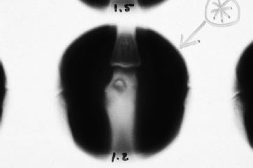
Plain radiographs reveal a small round lucency surrounded by sclerosis or a cortical reaction (Fig. 90–2). Lesions not demonstrated on plain films require a bone scan or computed tomography scan (Fig. 90–3). Bone scan may be necessary to demonstrate a sclerotic nidus (Fig. 90–4).
Differential Diagnosis
Ganglion
Enchondroma
Aneurysmal bone cyst
Giant cell tumor
Chondrosarcoma
Bone infection
Osteoid osteoma
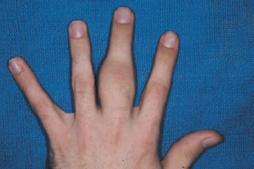
Figure 90–1. Clinical appearance of a large mass of the left long finger proximal phalanx.