Chapter 27 Orthoses and Insert Management of Common Foot and Ankle Problems
Forefoot
Intractable plantar keratosis (IPK)
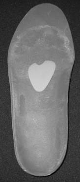
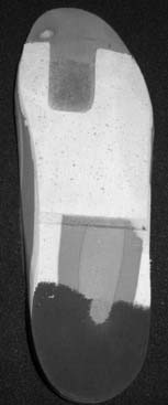
IPKs are calluses under bony prominences on the plantar aspect of the foot. They may be caused by a plantarflexed metatarsal head because of a hammertoe or a fracture, the elevation of an adjacent lesser toe metatarsal head that causes a transfer of pressure, or developmental problems of a similar nature (second metatarsal head callus adjacent to a bunion; a rotated fifth metatarsal head in a bunionette, a prominent sesamoid, and so forth). The solution is relatively simple: material is placed proximal to or around the prominent area (“posting”) to “offload” the prominent area and softer material is placed under the callus and prominence to cushion it. Using a material such as cork built into the insert material, we make a full-length, total-contact insert (TCI) with posting proximal to the lesion and create a well under the lesion. We then fill this well with a viscoelastic polymer, which adds excellent cushion, does not flow out of the well, and compresses more slowly than most other materials (Figs. 27-1 and 27-2). A similar solution is used for apical calluses (on the tips of hammer, claw, or mallet toes).
Morton’s neuroma or intermetatarsal neuritis
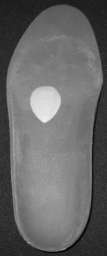
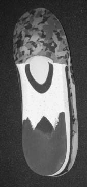
Irritation of the intermetatarsal nerve, which leads to neuritis or intraneural fibrosis (the Morton’s neuroma), is anatomically caused by the distal edge of the intermetatarsal ligament between the adjacent plantar plates of the metatarsophalangeal (MTP) joint. A metatarsal pad made of felt or other less compressible materials can be placed under the adjacent metatarsals proximal to the condylar heads of the adjacent metatarsals, thereby elevating them to decrease the contact of the edge of the plate when the patient is standing and walking. Some doctors place the pad on the patient’s foot with some adhesive or attach it to the sock liner in the shoe. We prefer to incorporate the pad into a total-contact insert, professionally placing the pad in the right place for the patient (Figs. 27-3 and 27-4). We also may add other features to the insert, such as longitudinal arch support when a symptomatic, flexible flatfoot accompanies the problem.
Ulcers under the metatarsal heads
Ulcers or deep blisters may occur under the metatarsal heads. This is a particularly common and challenging condition with the insensate foot but can occur in athletes as well. Although it is critical to analyze why the ulcer or blister occurred and to recognize the presence of structural problems, the pedorthic approach is an important adjunct to care. The insert should be full length, with posting around a relief well under the ulcer. Again, we fill this well with the viscoelastic polymer. In addition, a relief well also is created in the insole of the shoe by use of a burring tool. Finally, a mild rocker sole is placed on the outside of the shoe with the apex proximal to the ulcer site (Fig. 27-5). In the past, a metatarsal bar was placed in this location of the outer sole, but the rocker sole allows much easier walking than the bar. Before this stage of care, some surgeons may use total-contact casting, various commercially available boots that unweight the sole of the foot, and heel-weight-bearing–only postsurgical shoes. All of these measures may, at one stage or another, be adjunctive during the care of these problems. The orthoses and modified shoe may be used after the acute care to prevent later recurrence.
Metatarsopharangeal joint synovitis, “turf-toe,” arthritis, hallux rigidus, and rheumatoid arthritis
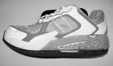
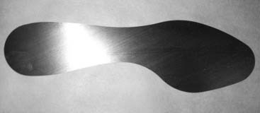
The treatment of an inflammatory condition of these joints should be immobilization while still allowing the patient to ambulate. This can be accomplished by using a stiff-sole shoe or insert. This effect can be obtained by placing a thin, spring-steel shank between the cushioned, total-contact insert and the insole on the shoe, or by incorporating the stiff material within the insert, or adding it to the sole of the shoe between the outer sole and midsole, or using a shoe that is made with a stiff shoe from the factory (Figs. 27-6 and 27-7). It is essential, however, to also use a rocker sole on the shoe (see Fig. 27-5) so that the patient can walk without the foot lifting up within the shoe; this would not only make the walking difficult but also increase the symptomatology. In a patient with hallux rigidus, there are two problems: pain in the joint from impingement, arthritis, and synovitis, and lack of motion. The previous prescription deals with these problems well, but some physicians will use the more rigid insert “Morton’s extension,” which lies from the heel to the end of the great toe but not all the way across the foot (Fig. 27-8).
< div class='tao-gold-member'>
Related posts:
 Impingement Syndromes of the Foot and Ankle
 Dermatologic, Infectious, and Nail Disorders
 Ankle Sprains, Ankle Instability, and Syndesmosis Injuries
 Assessment and Treatment of the Elite Athlete: Helpful Hints and Pertinent Pearls
 Ankle and Midfoot Fractures and Dislocations
 An International Perspective on the Foot and Ankle in Sports
Stay updated, free articles. Join our Telegram channel

Full access? Get Clinical Tree


