Chapter 11 Dermatologic, Infectious, and Nail Disorders
Dermatologic Disorders: Environmental/ Anatomic
Contact Dermatitis
Contact dermatitis (Fig. 11-1) is fairly common in an athletic population. Athletes encounter multiple exposures, including adhesive tape, compound of benzoin, topical medications (antibiotics, antifungals, and antiseptics), and rubber-containing sports equipment. Some athletes even have reactions to the leather products of which most sports shoes are composed. Contact dermatitis presents as an inflammatory response of the skin to an offending irritant. The main pathologic feature of contact dermatitis is intracellular edema of the epidermis, resulting in intraepidermal vesicle and bullae formation in acute cases. In chronic cases, a presentation with papules, scarring, and lichenification can be seen.1 Allergic contact dermatitis will be seen in individuals who previously have been sensitized to the allergen. A delayed hypersensitivity reaction will be seen over the course of several hours. In athletes, contact dermatitis is seen most commonly on the dorsum of the foot and toes. Fisher2 stressed that the moist environment within the athletic shoe is a major component in the development of contact dermatitis. It was stated that feet that were kept dry would not develop this form of dermatitis.
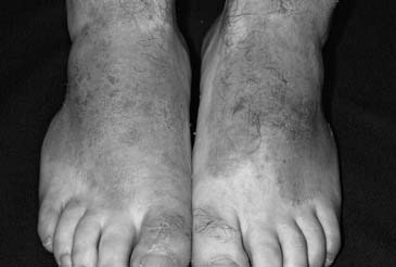
Figure 11-1 Contact dermatitis.
(From Habif, Clinical dermatology, St Louis, 2004, Mosby: p. 92, Figure 4-16.)
 Pearl
PearlFrostbite
Injury induced by cold exposure has been recognized for thousands of years.3 Frostbite involves the skin and potentially the soft tissues of the foot and ankle in athletes that are exposed to prolonged cold environments. Injury can occur, however, even with brief exposure of an unprotected foot to a cold, conductive surface (metal, concrete). Several terms are used to describe this phenomenon, including frostnip, chilblains, and frostbite. Frostnip is described as nonfreezing injury to the skin tissues that can commonly be seen in the toes. Associated symptoms include numbness and tingling. Cellular injury is absent in frostnip. Chilblains is associated with a more significant nonfreezing cold injury seen at temperatures below 59 °F in which mild tissue damage is seen in the form of minor vascular injury and tissue swelling. Frostbite is the destruction of body tissues because of freezing (below 32 °F) and ice crystal formation in the tissues, which causes cell lysis and tissue destruction. The tissue damage seen in association with frostbite is caused by two distinct mechanisms. First, ice crystal formation in the intracellular space leads to cellular dehydration and destruction. Second, damage to the vascular endothelium leads to inefficient delivery of blood to the injured tissues, further complicated by edema and swelling. Ultimately, further cell deterioration is seen secondary to hypoxia.4
The diagnosis of frostbite is made on the basis of history and physical examination. It is difficult to predict the degree of tissue injury for weeks following the exposure. With severe injury, it may take months before a clear delineation of viable versus nonviable tissue can be made. There is no current radiologic technique that can reliably distinguish the line of demarcation of injured tissues in the immediate postinjury period. Continued research using technetium scintigraphy and magnetic resonance techniques is needed to identify whether any available radiographic procedure may allow for early distinction of viable tissues.4
The treatment of frostbite injuries can be divided into three phases, including initial evaluation, acute care, and long-term follow-up. McCauley et al.5 described a treatment protocol for frostbite. This protocol can be applied to active individuals who suffer injuries to the lower extremity. Initially, the athlete should be admitted for rapid rewarming of the affected area in warm water (104 ° to 108 °F for 15 to 30 minutes or until thawing has completed). After the completion of rewarming, the affected parts should be treated as follows: white blisters should be debrided and topical treatment with aloe vera should be applied every 6 hours. Hemorrhagic blisters should be left intact, with topical aloe vera administration every 6 hours. The patient’s lower extremities should be elevated and splinted as needed. The athlete should be given antitetanus prophylaxis. Regular administration of anti-inflammatory medications is recommended. Analgesia should be accomplished using narcotic medications. Antibiotic coverage should be started and continued for the first 2 to 3 days or until signs of superinfection have cleared. Daily hydrotherapy should be implemented for 30 to 45 minutes at a temperature of 104 °F. Lastly, the patient should avoid smoking during this time to prevent peripheral vasoconstriction. When evaluating and treating patients with frostbite, the clinician should avoid rubbing the involved region because this can cause additional damage to the injured tissue. It also is important to ensure that there is not a possibility of refreezing after the rewarming process has taken place. If the potential for refreezing exists, rewarming should be delayed until further cold injury can be avoided. If refreezing does occur, significantly more tissue injury can manifest. Most athletes with frostbite injuries of the foot and ankle will present with findings consistent with a superficial injury. After rewarming, the athlete can be treated in an outpatient setting using appropriate topical formulations and analgesics. Close follow-up is necessary.
 Pearl
PearlHyperhidrosis
The treatment for hyperhidrosis can be challenging. Topical agents such as glutaraldehyde solution can be administered in an attempt to reduce perspiration through the denaturation of keratin with resultant occlusion of the pores of the sweat glands. Aluminum compounds such as aluminum chloride function as antiperspirants and can be used topically, as well. Darrigrand et al.6 studied the application of antiperspirants to the feet of cadets in an attempt to decrease foot sweat accumulation and injuries. They demonstrated a 50% decrease in foot sweat accumulation and a reduced occurrence of trench foot and friction blisters. There was, however, an increased incidence of contact dermatitis. Oral administration of anticholinergic agents such as propantheline, glycopyrrolate, benztropine, and oxybutynin also has been advocated. In addition, neuromuscular blocking agents such as botulinum toxin can be used to inhibit transmission of nerve impulses at the neuromuscular junction of skeletal muscle and/or the autonomic ganglia. It is recommended to perform a nerve block of the posterior tibial nerve and the sural nerve before to botulinum toxin treatment for plantar hyperhidrosis.7
 Pearl
PearlHyperkeratosis
Hyperkeratosis (Fig. 11-2) in the form of corns and calluses is a standard feature for many athletes. These lesions are produced as pressure and friction are applied repeatedly to the skin overlying the osseous structures. In an attempt to protect from skin breakdown, the body produces these regions of hyperkeratotic tissue. Many athletes who have symptoms related to their hyperkeratosis complain of localized pain and discomfort. Interestingly, the hyperkeratotic tissue itself is not what causes the pain, but rather underlying bursitis and nerve irritation. The most common types of hyperkeratosis include helomas, tylomas, and intractable plantar keratoma. Helomas are synonymous with corns and usually are found on the toes. Tylomas also are known as calluses and they usually are found over bony prominences, especially in the region of the metatarsal heads. Intractable plantar keratoma is defined as a cone-shaped keratotic plug within a tyloma.
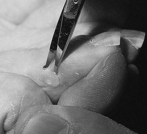
Figure 11-2 Hyperkeratosis (corn).
(From Habif, Clinical dermatology, St Louis, 2004, Mosby: p. 928, Figure 27-5.)
Calluses are seen almost universally on the feet of athletes. As described earlier, this is the body’s response to wear and tear. Many times, athletes are protected from skin breakdown and potential infection by this hyperkeratotic process. Callus formation becomes problematic when there is an overproliferation of the keratin tissue leading to underlying bursitis, neuroma, or neuritis.8 Intractable plantar keratoma is a highly painful lesion in the region of the plantar aspect of the metatarsal heads. Intractable plantar keratoma can be extensive and symptomatic enough to affect an athlete’s performance.9 Deformities of the metatarsophalangeal joint that produce increased plantarflexion in this area lead to the development of intractable plantar keratoma in the metatarsal head regions receiving the most plantar pressure.
 Pearl
PearlXerosis
 Pearl
PearlRelated posts:

Stay updated, free articles. Join our Telegram channel

Full access? Get Clinical Tree


