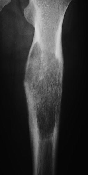
Fig. 1
Anteroposterior radiograph of the distal femur showing a destructive lesion in the lateral aspect of the metaphysis. Note the bone destruction and periosteal reaction. This appearance is very typical of osteosarcoma
Staging evaluation – a magnetic resonance imaging of the lesion determines the size of the lesion and the absence or presence of discontinuous tumor (skip metastases). A computerized tomography scan of the chest is used to look for pulmonary metastases (appears as one or more nodules), and a technetium bone scan is used to search for bone metastases.
Treatment – the preoperative chemotherapy is followed by wide resection of the tumor and reconstruction. Most patients receive postoperative chemotherapy for 6–12 months. Patients who present with localized disease (Stage II) have a 60–70 % long-term survival, while patients who present with pulmonary nodules (Stage IV disease) have a survival of approximately 25 %.
Ewing’s tumor – this is the second most common cause of a malignant bone sarcoma in the young patient. In addition to presenting with pain and a soft tissue mass, patients may also have constitutional symptoms such as fever and fatigue. The erythrocyte sedimentation rate and white blood cell count may be elevated. When the radiographs and the systemic manifestations are considered, osteomyelitis is often considered higher in the differential diagnosis than Ewing’s tumor.
Imaging – the radiographs often show a destructive lesion (Fig. 2). The magnetic resonance imaging scan often shows a large soft tissue mass, and there may be a high level of periosteal reaction.


