Fig. 31.1
Example of a hybrid SPECT/CT system consisting of a 16-slice CT and a dual-head gamma camera
SPECT/CT is a hybrid imaging modality that combines a 3D scintigraphy (SPECT) with a multi-slice computerised tomography (CT).
Our patients receive a commercial 500–700 MBq, 99mTc-marked diphosphonate (e.g. MDP, HDP) injection. Planar scintigraphic images are taken in three phases, the perfusion phase (immediately after injection), the blood pool phase (from 1 to 5 min after injection) and the delayed metabolic phase (from 2 h after injection, Fig. 31.2).
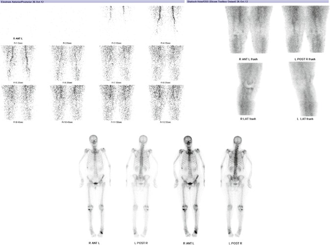
Fig. 31.2
Planar scintigraphic images are taken in three phases, the perfusion phase (immediately after injection), the blood pool phase (from 1 to 5 min after injection) and the delayed metabolic phase (from 2 h after injection)
SPECT/CT is performed with a matrix size of 128 × 128, an angle step of 32 and a time per frame of 25 s 2 h after injection. For the CT, a specific protocol is used that takes 3 mm slices of the femoral head, 0.75 mm slices of the knee joint (30 cm above and below) and 3 mm slices of the ankle joint (Fig. 31.3). The whole TKR and parts of the femoral and tibial shaft, particularly important in the case of stems, have to be in the field of scanning. The protocol used was modified according to the Imperial Knee Protocol, which is a low-dose CT protocol [4].
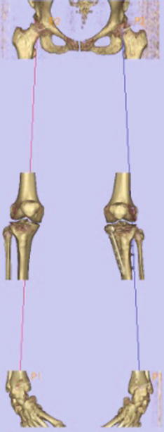
Fig. 31.3
Specific SPECT/CT scanning protocol including 3 mm slices of the femoral head, 0.75 mm slices of the knee joint (30 cm above and below) and 3 mm slices of the ankle joint
In the era of planar scintigraphy and SPECT, increased tracer uptake in the perfusion, blood pool and delayed phase of three-phase scintigraphy was indicative of an infection in patients after knee replacement [2]. Aseptic loosening was generally diagnosed if the delayed phase showed increased tracer uptake around the prosthesis (e.g. stem), while there was none in the negative perfusion and blood pool phase [2].
With the introduction of SPECT/CT, these diagnostic guidelines and rules seem to change as SPECT/CT offers a richer source of combined mechanical, structural and metabolic information of the patient’s knee joint. The metal artefact splatter, which is an inherent problem of CT and impairs the assessment of the prosthetic-bone interface, is reduced [2, 6, 7, 9]. For the first time, a combined assessment of bone metabolism (including remodelling around the prosthesis), mechanical alignment, component position and structural changes is possible. In particular, the combination of 3D analysis of component position, mechanical and anatomical axes as well as distribution and intensity of SPECT tracer uptake values is beneficial for the orthopaedic surgeon [6, 7, 9–11].
SPECT/CT offers a richer source of combined mechanical, structural and metabolic information of the patient’s knee joint. The metal artefact splatter, which is an inherent problem of CT and impairs the assessment of the prosthetic-bone interface, is reduced.
In our own study, we investigated the position of TKR components on standard radiographs and 2D axial CT images as well as on 3D CT reconstructions [8]. Plain radiographs were highly reliable at measuring the tibial slope but showed wide variability for all other measurements. 2D CT also showed wide variability. 3D CT was highly reliable, even when measuring rotation of the femoral components and significantly better than 2D CT [8]. The conclusion was that, from now on, 3D CT should be the investigation of choice for determination of TKR component position [8] (see Chap. 29).
For the evaluation of SPECT tracer uptake, we have introduced and validated a standardised, easily applicable and reliable algorithm using SPECT/CT in patients after TKR [7]. The localisation scheme differentiates the knee into 9 tibial, 9 femoral and 4 patellar regions on standardised axial, coronal and sagittal slices [7]. Along with the introduction of a standardised localisation scheme, pathology-specific distribution patterns and intensity thresholds of SPECT/CT tracer uptake could be identified, which then reflect mechanical loosening, instability, component malposition or patellofemoral problems [7].
In a first study we piloted, the validated SPECT/CT algorithm including the 3D analysis of component position and tracer uptake assessment on a series of 23 knees with pain after TKR [9]. SPECT/CT significantly changed the diagnosis and treatment proposed, independently of the previous intention to revise or treat the patient nonsurgically [9]. Within this series progression of patellofemoral osteoarthritis within the first 5 years after primary TKR, tibial or femoral loosening or oversizing of TKR components were most frequently reported [9]. Patients with femoral mechanical loosening presented with increased tracer activity around the femoral tray [9]. Patients with externally malrotated tibial tray showed significantly higher tracer activity in the medial patellar facet and by trend in the femur [9]. The posterior tibial slope <3° or >10° was associated with increased femoral tracer uptake [9]. Patients with patellofemoral OA as leading cause for their knee pain showed significantly higher tracer uptake in the patella than others [9]. Patients with loosening of the femoral component showed significantly higher tracer uptake in the tibia, which also extended to the prosthetic interface [9]. The femoral uptake was also higher but did not reach statistical significance [9].
In currently unpublished data, we have extended our work to a series of 500 patients with painful TKR. Along with the 3D analysis of tibial and femoral component position (varus-valgus, flexion-extension, internal and external rotation), the knee was evaluated with regard to areas of increased tracer uptake (“hotspots”). With this new study, we aimed to identify the typical pattern of tracer uptake distribution and intensity values in patients after total knee replacement. The above findings were correlated with the type of TKR, the time from primary TKR surgery, cemented or uncemented arthroplasty and intraoperative findings at revision surgery (loose vs. well-fixed TKR components). The results will be published in detail in a peer-reviewed journal. From this study, we recommend consideration of the age of TKR, type and design of TKR, the mode of fixation (cemented vs. uncemented) and the mechanical alignment when analysing SPECT/CT in patients after TKR. All these factors significantly influence the bone tracer uptake pattern.
In contrast, there is only scarce evidence about the use of SPECT/CT diagnosing infection after knee replacement [3, 11, 1]. However, these few studies showed a significant increase of specificity and sensitivity in diagnosing infection after knee replacement [3, 11, 1]. The sensitivity was 0.89, the specificity was 0.73 and the positive and negative predictive values were 0.57 and 0.94, respectively, [1].
Growing clinical evidence on the use of SPECT/CT in painful TKR helps to establish diagnosis and guide further treatment. In contrast, there is only scarce evidence about the use of SPECT/CT in infection after TKR.
One of the biggest barriers to the use of SPECT/CT has been its lack of availability. SPECT/CT was only available in university hospitals. With improved availability, the use of SPECT/CT seems likely to expand in the coming years. Another reason that SPECT/CT has not gained introduction in diagnostic guidelines and routine daily practice yet has been the insufficient analysis tools available.
Since Hirschmann et al. presented a novel method of standardised volumetric 3D analysis of SPECT/CT imaging using a customised specific software solution (Orthoexpert©; also see Chap. 7), this limitation has been overcome [10]. Clearly, the authors demonstrated a method to quantitatively and volumetrically measure the intensity of SPECT tracer uptake in any area of the knee joint, a method to localise SPECT/CT data in 3D using clinically relevant anatomical landmarks and frames of reference, a method to normalise orthopaedic SPECT/CT data and a method of thresholding SPECT/CT data, which facilitates distinguishing clinically relevant hot spots from background activity [10].
In summary, SPECT/CT is a promising imaging modality in patients after knee replacement as it offers the combined analysis of tracer uptake distribution and intensity as well as the prosthetic component position and the evaluation of mechanical or anatomical alignment into one imaging modality. All these factors are known to be important for outcome after knee replacement. A combined analysis offers new dimensions of diagnostics such as investigating the influence of alignment or prosthetic component position on tracer uptake distribution or intensity. Having a broader knowledge about the mutual influences of the above described factors on SPECT tracer uptake will in the future lead to new diagnostic algorithms, thus improving the specificity and sensitivity of diagnosing aseptic or septic loosening in patients after knee replacement. SPECT/CT seems to be the one and only imaging modality that allows one to assess the patient’s biomechanics. Hence, it can be called the biomechanical eye of the orthopaedic surgeon.
Related posts:
 Diagnosis of Periprosthetic Joint Infection After Total Knee Replacement
How Can Preoperative Planning Prevent Occurrence of a Painful Total Knee Replacement?
Causes and Diagnosis of Aseptic Loosening After Total Knee Replacement
Discussion to Chap. 16: Mid-Flexion Instability After TKR Due to Femoral Malrotation
Specific Orthopaedic Imaging Analysis Software: Clinical Benefit for TKR Revision Surgeon
Constrained Condylar Total Knee Replacement
Diagnosis of Periprosthetic Joint Infection After Total Knee Replacement
How Can Preoperative Planning Prevent Occurrence of a Painful Total Knee Replacement?
Causes and Diagnosis of Aseptic Loosening After Total Knee Replacement
Discussion to Chap. 16: Mid-Flexion Instability After TKR Due to Femoral Malrotation
Specific Orthopaedic Imaging Analysis Software: Clinical Benefit for TKR Revision Surgeon
Constrained Condylar Total Knee Replacement
Stay updated, free articles. Join our Telegram channel

Full access? Get Clinical Tree


