Fig. 6.1
Schematic drawing of a shoulder joint, before and after insertion of a Delta III prosthesis. The center of rotation (red dot) is medialized, but not distalized. This created a mechanical conflict between the inferior part of the humeral cup and the scapular neck
This mechanical conflict not only decreased the stability of the humeral component in adduction but also resulted in a bony defect or notch at the inferior part of the scapular neck. Such a notch was a common radiographic finding in patients treated with a Delta III or equivalent reverse shoulder prosthesis [2, 3, 4]. It appeared within the first months after the operation and was associated with an abrasion of the inferior part of the polyethylene liner [5]. We hypothesized that this mechanical conflict and the related complications can be avoided if the glenoid component is fixed more inferiorly on the glenoid bone.
Materials and Methods
Eight fresh-frozen shoulder specimens were used for this study. [6] After thawing, all soft tissues were removed, except the coracoacromial ligament. A Delta III humeral prosthesis with a lateralized cup was inserted into the humeral shaft in normal retroversion. The scapula was fixed in a vice with the scapular blade and the glenoid surface oriented vertically. A glenoid component with a diameter of 36 mm was attached to the glenoid in the following positions (Fig. 6.2): glenosphere centered on the glenoid, leaving the inferior part of the glenoid bone uncovered (configuration A), glenosphere flush with the inferior glenoid rim (configuration B), glenoid baseplate (metaglene) flush with the glenoid rim and glenosphere extending beyond the inferior border of the bone (configuration C), and glenosphere tilted downward 15° and flush with the inferior border of the glenoid (configuration D). The humerus was passively abducted and adducted in the scapular plane, in the frontal plane and in the sagittal plane. Movements were stopped as soon as the humeral head or the humeral cup came into contact with the bone of the scapula. The corresponding glenohumeral abduction and adduction angles were recorded.
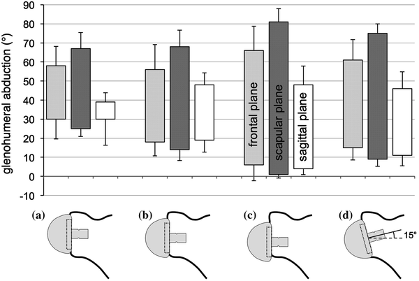

Fig. 6.2
Graph showing the different configurations of the glenosphere and the corresponding maximum abduction and adduction angles in the frontal, scapular, and sagittal plane
Results
The results of the glenohumeral abduction and adduction angle measurements are shown in Fig. 6.2. In all cases, range of motion was better in the scapular plane than that in the frontal or sagittal plane. The highest abduction and the best adduction angles were measured when the glenosphere extended beyond the inferior glenoid rim (test configuration C). Insertion of the glenoid component with a slightly inferior tilt decreased the surface area of the glenoid bone and brought the humerus closer to the scapular pillar.
Discussion
Clinical and other experimental studies confirmed these results [7–9], and efforts were made to reduce the risk of notching. Some companies reversed the materials and manufactured a polyethylene glenosphere and a metallic cup (Affinis reversed, Mathys Ltd., Bettlach, Switzerland; SMR Lima, Villanova di San Daniele del Friuli, Italy). This modification did not solve the mechanical conflict, but it significantly reduced the incidence and the severity of radiologic notching [10]. This confirms that a scapular notch is rather a consequence of a polyethylene wear induced osteolysis than the result of a direct abrasion of the bone [5].
Some companies designed eccentric or bigger glenospheres in order to increase the inferior offset and others lateralized the center of rotation [11, 12]. The Duocentric prosthesis (Aston Medical, Saint-Etienne, France), which was developed by the engineers of the Delta III prosthesis and the successors of Grammont, combines these two modifications and has in addition a 10° smaller neck–shaft angle. These modifications seem to be effective. The first 100 patients treated with such a prostheses in our clinic have not developed a notch to date (Fig. 6.3).
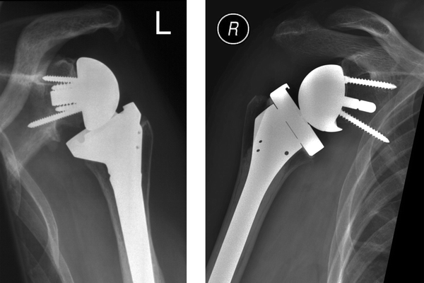

Fig. 6.3
Typical anteroposterior radiograph of a Delta III prosthesis with a scapular notch (left side) and a Duocentric prosthesis without a scapular encroachment (right side). Both prostheses have a 36-mm glenosphere, but the position of the center of rotation and the neck-shaft angle are different (see Table 6.1)
Influence of Prosthesis Design on Internal and External Rotation
Similar to the superior liftoff of the polyethylene cup in adduction, an anterior liftoff can be observed intraoperatively during external rotation of the adducted arm. It is the result of an impingement of the postero-inferior part of the humeral cup with the posterior aspect of the scapular neck. The angle, at which this phenomenon occurs, depends on the geometry of the prosthesis and the retroversion of the humeral component. It is particularly small, if a Grammont-type prosthesis with a 36-mm glenosphere and a neck–shaft angle of 155° is used. Patients treated with such prosthesis are often unable to externally rotate their arm more than 10° (Fig. 6.4).
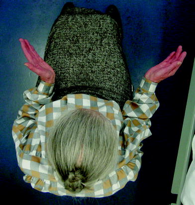

Fig. 6.4
Maximum external rotation with the arm at the side 2 years after insertion of a Delta III prosthesis on the left side and a Delta X-tend prosthesis on the right side
Materials and Methods
We quantified the internal and external rotation angles of different types and sizes of reverse shoulder prostheses available on the European market. The prostheses selected for this study were the Delta III prosthesis (DePuy International Ltd, Leeds, England) as the gold standard, the Anatomical Inverse/Reverse prosthesis (Zimmer, Inc., Warsaw, USA), the Affinis Inverse (Mathys Ltd., Bettlach, Switzerland), and the Duocentric prosthesis (Aston Medical, Saint-Etienne, France). The geometric parameters of these prostheses are listed in Table 6.1. In order to eliminate any possible anatomical difference between specimens, the tests were made on identical synthetic right shoulder models. The shape of these models corresponded to the shape of adult human bones. The glenoid baseplates were fixed in such a way that the inferior contour was flush with the inferior glenoid rim. That ensured a small overhang of the glenosphere. The humeral components were inserted into the synthetic bones with 10° of retroversion. An aluminum rod was placed in the distal part of the medullary canal and represented the rotational axis of the humerus. The forearm was replaced by a 2.5-mm Kirschner wire, in 90° of elbow flexion. The scapula was rigidly fixed to a custom-made metallic frame, with the scapular blade and the glenoid fossa oriented vertically. The humerus was centered on the glenosphere, and the aluminum rod was attached to an arc, which was placed in the scapular plane. The arm was then passively internally and externally rotated until the humeral cup came into contact with the scapular neck. The position of the arm was recorded with a MicroScripe 3-D digitizer (Solution Technologies, Inc., Oella, USA), and the corresponding abduction and rotation angles were calculated using vector geometry. These measurements were repeated at different angles of abduction in the scapular plane. A 4th-order polynomial was fitted to the measured values.
Table 6.1
Geometric parameters of the different reverse prostheses tested. The lateral offset refers to the surface of the glenoid bone
Diameter of glenosphere (mm) | Inferior overhang of glenosphere (mm) | Lateral offset of center of rotation (mm) | Depth of the cup (mm) | Neck-shaft angle (°) | |
|---|---|---|---|---|---|
Delta III | 36 | 3.5 | 0 | 8.0 | 155 |
Anatomical 36 | 36 | 1.0 | 6.0 | 8.0 | 155 |
Anatomical 40 | 40 | 5.0 | 6.0 | 8.0 | 155 |
Affinis 36 | 36 | 4.5 | 1.7 | 8.5 | 155 |
Affinis 42 | 42 | 7.5 | 1.7 | 10.0 | 155 |
Duocentric 36 | 36 | 4.0 | 5.0 | 6.0 | 145 |
Results
The results are shown in Fig. 6.5. Neutral rotation was defined as the position, in which the forearm was perpendicular to the scapular plane, that means horizontal. In vivo, neutral rotation of the adducted arm is normally defined as the position, in which the forearm is perpendicular to the frontal plane. Because the scapular plane and the frontal plane form an angle of about 30°, neutral rotation of the adducted arm in our experiments corresponds to about 30° of internal rotation in vivo.
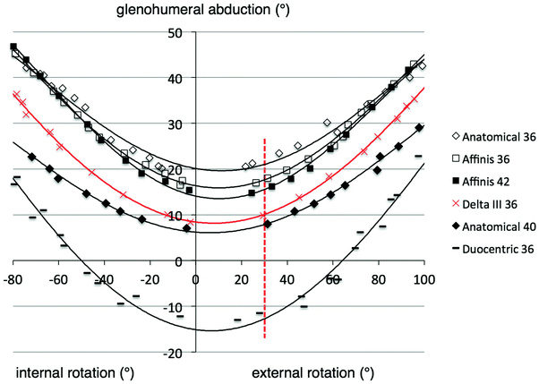

Fig. 6.5
Graph showing maximum internal and external rotation of different shoulder prostheses tested in a plastic shoulder model. The measurements were made at different glenohumeral abduction in the scapular plane. Neutral rotation was defined as the position, in which the forearm was perpendicular to the scapular plane. Neutral rotation of the adducted arm in vivo corresponds to about 30° of external rotation (dotted red line) in our experiments
The lowest abduction angles were obtained in 10° of external rotation, which corresponded to the retroversion of the humeral component. The rotation amplitude increased with increasing glenohumeral abduction. There were significant differences between the different types of reverse prostheses. The worst results were obtained with the 36-mm Anatomical prosthesis. The glenoid plate (metaglene) of this prosthesis is relatively voluminous and the 36-mm glenosphere overlaps the inferior border of this plate only 1 mm. Adduction as well as internal and external rotation was much better when the 40-mm glenosphere was fixed on the same glenoid plate. This was not the case for the Affinis prosthesis. The rotation amplitudes of the Affinis prosthesis did not depend on the diameter of the glenosphere. The reason is that the bigger glenosphere of the Affinis prosthesis is combined with a deeper cup. If the depth of the cup increases proportionally with the diameter of the glenosphere, the arc of enclosure does not change. The best rotational amplitudes were obtained with the Duocentric prosthesis.
Discussion
Internal and external rotation was stopped when the humeral cup abutted against the scapular neck. That means that the morphology of the scapula influences the range of motion of a reverse prosthesis. The values presented in Fig. 6.5 are therefore only valid for the shoulder model tested in this study. Range of motion may be better, if the scapular neck is longer, or worse, if the scapular neck is shorter than in our plastic model.
The maximum internal and external rotation angles depend on the retroversion of the humeral component. More retroversion improves external rotation at the expense of internal rotation, and vice versa. The choice of retroversion is therefore particularly important if prostheses with small rotation amplitudes are used.
The measurements were made passively and without any soft tissues. In vivo, glenohumeral motion may be smaller, either due to muscular weakness or due to soft tissue impingement. The inferior joint capsule and the long head of the triceps muscle may restrict adduction, the conjoint tendon may hinder internal rotation, and a stiff subscapularis may limit external rotation.
An interesting finding of this study was that a bigger glenosphere does not necessarily mean a better range of motion. The impingement-free range of motion depends among other factors on the relationship between the diameter of the glenosphere (D) and depth of the cup (d). The arc that a cup encloses on a sphere can be calculated with the formula: α = 2 arccos (1 − 2d/D). [13] The bigger the arc of enclosure, the smaller the impingement-free range of motion.
The Duocentric prosthesis performed much better than the original prosthesis of Grammont. The reasons therefore are the lateral and inferior offset of the glenosphere and the 10° lower neck–shaft angle of the humeral implant. This study does not allow quantifying the effect of each single parameter. But the differences in range of motion between these two prostheses can also be observed during surgery and during clinical examination of the patients (Figs. 6.4 and 6.6).
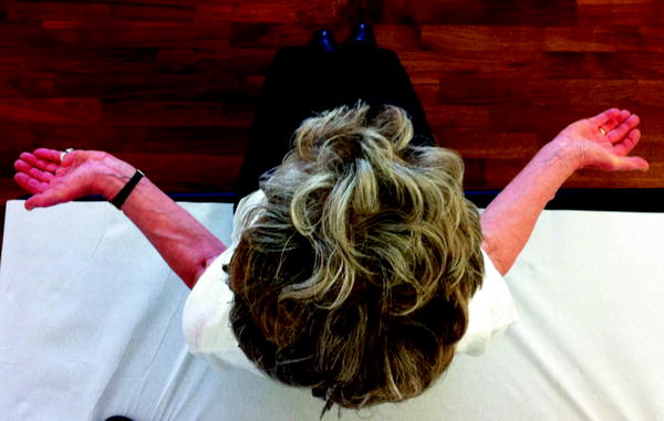

Fig. 6.6
Maximum external rotation with the arm at the side 1 year after implantation of Duocentric prostheses on both sides
Shoulder Kinematics in Vivo
Most patients treated with a reverse shoulder prosthesis are able to elevate their arm above the horizontal line. Full elevation, however, is usually not possible. Elevation of the arm is a combination of glenohumeral and thoracoscapular motion. The relationship between these two movements is called shoulder rhythm and is about 2:1 in normal shoulders, meaning that 2/3 of total motion occurs in the glenohumeral joint and 1/3 in the thoracoscapular gliding space. A reduction of global elevation can therefore be due to a reduction of glenohumeral motion, thoracoscapular motion, or both of them.
We determined the shoulder rhythm of 10 patients [14] treated either with a Delta III (DePuy International Ltd, Leeds, England) or an Aequalis reversed prosthesis (Tornier, Montbonnot, France). The geometric parameters of these two prostheses were identical (see Table 6.1). All patients had enough strength to actively elevate their arm above the shoulder level. There were two men and eight women with an average age at the time of surgery of 71 years (range 53–84 years). Indications for reverse shoulder arthroplasty were a cuff tear arthropathy (4), a failure after hemiarthroplasty (4), a failure after osteosynthesis of a proximal humerus fracture (1), and an acute fracture (1). Six patients had one to three previous operations.
A deltopectoral approach was made, and a 36-mm glenosphere was used in all patients. The humeral component was inserted into the shaft in 0°–20° of retroversion. The patients were reviewed at a mean follow-up of 26 months. The assessment consisted of a structured interview, a clinical examination with photographic documentation, and a radiographic evaluation. For the purpose of this study, true anteroposterior radiographs were made with the arm successively held in four different angles of abduction. Therefore, the patients were standing in front of the screening board, slightly oblique with the examined scapula applied against the X-ray cassette. In contrast to the standard examination technique, the X-ray beam was not inclined but oriented horizontally in order to limit projection errors that would adversely affect the angular measurements. The first radiograph was taken with the arm held in adduction and neutral rotation. The patients were then asked to actively elevate their arm in the scapular plane (parallel to the X-ray film) to above the shoulder level and to maintain this position until the second radiograph was made. Two additional radiographs were taken with the arm in an intermediate position of about fifty and seventy-five degrees of abduction, respectively.
Stay updated, free articles. Join our Telegram channel

Full access? Get Clinical Tree


