Fig. 1
Patient with midfoot arthritis primarily at the middle column. Note the subtle asymmetric narrowing on the AP radiograph. The presence of 2nd TMT degeneration is clearly seen on the lateral radiograph (arrow)
Subchondral sclerosis/cysts
Osteophytes and bony erosions – best seen on lateral view
Disruption of Meary’s line (loss of colinearity between talus and 1st MT) with apex deformity at midfoot and longitudinal arch collapse on lateral view
Forefoot abduction deformity. Critical to differentiate from PTTD flatfoot deformity where TN in abduction > TMT (Fig. 2)
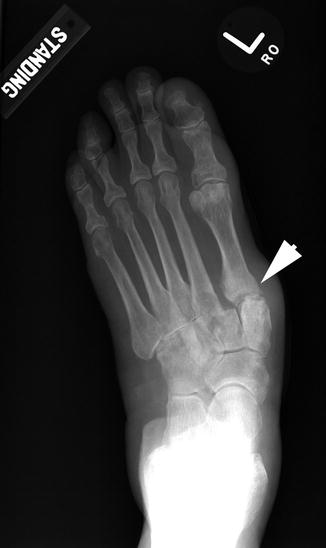
Fig. 2
AP WB radiograph demonstrating abduction deformity of the foot that is clearly focused at the TMT joints (arrowhead). Note the minimal amount of TN joint subluxation despite the severe deformity, indicating this is a midfoot driven flatfoot and not related to PTTD
Treatment
Nonoperative
A stiff carbon fiber plate inserted underneath the insole of the shoe or a rocker-bottom shoe will offload stress on the midfoot during gait and may relieve mild degenerative symptoms.
Rigid deformity should be managed with accommodative, not corrective orthotics that support and offload the deformity.
Corticosteroid/anesthetic injections: both diagnostic (determine symptomatic joints) and therapeutic
Operative
Indication is failure of nonoperative with persistent pain and disability. Arthrodesis is the treatment of choice. Goal of surgery is to obtain solid fusion and restoration of normal foot alignment.
Medial and middle column arthrodesis ensuring a normal anatomic alignment. Residual deformity will predictably result in a poor outcome (Fig. 3).
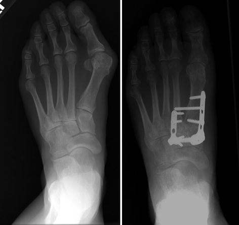
Fig. 3
A Lapidus and 2nd TMT arthrodesis was performed in this patient who presented with complaints of hallux valgus and midfoot pain with radiographic evidence of midfoot DJD. The patient had osteopenic bone and therefore plate fixation was chosen
Interpositional arthroplasty (motion sparring) of lateral column is preferred for symptomatic 4th and 5th TMT arthritis given the biomechanical advantage. However, no significant data is present to support motion-sparing procedures over arthrodesis.
A simultaneous gastrocnemius recession and/or hindfoot osteotomy might be required to restore normal foot alignment (Fig. 4).
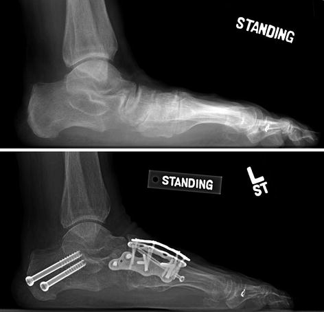
Fig. 4
Extended midfoot arthrodesis with concomitant medial slide calcaneal osteotomy was required in this patient who presented with midfoot arthritis and secondary hindfoot valgus deformity. Note the restoration of the longitudinal arch in the postoperative radiograph
Complications
Nonunion/malunion
Wound complications
Bibliography
1.
Nemec SA, Habbu RA, Anderson JG, Bohay DR. Outcomes following midfoot arthrodesis for primary arthritis. Foot Ankle Int. 2011;32(4):355–61.
2.
Patel A, Rao S, Nawoczenski D, Flemister AS, DiGiovanni B, Baumhauer JF. Midfoot arthritis. J Am Acad Orthop Surg. 2010;18(7):417–25.
3.
Russell DF, Ferdinand RD. Review of the evidence: surgical management of 4th and 5th tarsometatarsal joint osteoarthritis. Foot Ankle Surg. 2013;19(4):207–11.
4.
Verhoeven N, Vandeputte G. Midfoot arthritis: diagnosis and treatment. Foot Ankle Surg. 2012;18(4):255–62.
5.
Zonno AJ, Myerson MS. Surgical correction of midfoot arthritis with and without deformity. Foot Ankle Clin. 2011;16(1):35–47.
2 Kohler’s Disease
Take-Home Message
Pediatric avascular necrosis of the navicular of unknown etiology.
Boys preferentially affected.
25 % can be bilateral.
Usually self-limiting with favorable prognosis, surgery is not indicated.
Immobilization with short leg walking cast is indicated for activity-related symptoms.
Immobilization decreases duration of symptoms.
Definition
Avascular necrosis of the navicular bone of unclear etiology. Occurs in pediatric population. Patients usually present with pain (dorsomedial), swelling, and warmth/redness and limp.
However, some patients are asymptomatic.
More common in boys (up 80 % of cases).
25 % of cases are bilateral.
Etiology
Unknown
Pathophysiology
Blood supply: the central 1/3 of the navicular is a watershed area → avascular necrosis
Late ossification → predisposition to injury and mechanical compression
Radiography
Foot X-ray
Active disease: sclerosis, fragmentation, and flattening of navicular
Post resolution: bone remodeling and reconstitution, asymptomatic deformity can persist
Treatment
Usually self-limiting with good prognosis. Symptoms usually associated with activities.
Nonoperative
NSAIDs, activity modification (eliminate impact)
Shoe insert orthotics (accommodative)
Immobilization with short leg walking cast
Indication: activity-related pain and failure of above modalities
Operative
No indication for surgery
Complications
Very good prognosis without post-resolution sequela. Deformity is usually asymptomatic.
Bibliography
1.
Borges JL, Guille JT, Bowen JR. Köhler’s bone disease of the tarsal navicular. J Pediatr Orthop. 1995;15(5):596–8.
2.
DiGiovanni CW, Patel A, Calfee R, Nickisch F. Osteonecrosis in the foot. J Am Acad Orthop Surg. 2007;15(4):208–17.
3.


Tsirikos AI, Riddle EC, Kruse R. Bilateral Köhler’s disease in identical twins. Clin Orthop Relat Res. 2003;409:195–8.
Related posts:

Stay updated, free articles. Join our Telegram channel
Full access? Get Clinical Tree
