Mesenchymal Chondrosarcoma
Mesenchymal chondrosarcoma was first described by Lichtenstein and Bernstein in 1959. The other unusual chondroid tumors described by these authors do not have counterparts that we have recognized in the material available for study. If any lesions are present, they have been classed with chondrosarcoma, chondromyxoid fibroma, and chondroblastoma.
Primitive multipotential primary sarcoma of bone, as described in 1966, includes lesions that fit the descriptions of mesenchymal chondrosarcoma. Polyhistioma of bone and soft tissue is a more recently suggested term for malignant tumors that have areas of small round-to-oval cells. In such zones, the tumors bear a striking similarity to Ewing sarcoma, but they differ from Ewing sarcoma in that the other areas show various types of differentiation, especially chondroid, osteoid, or fibromatoid, and with corresponding matrices. Hence, mesenchymal chondrosarcoma may artificially exclude some related tumors. Unfortunately, the exact delimitations of these other varieties of so-called polyhistioma are not yet well defined.
INCIDENCE
The total of 48 (46 patients) mesenchymal chondrosarcomas in the Mayo Clinic series indicates the rarity of this lesion (Fig. 7.1). They accounted for about 0.7% of malignant tumors. Included in this total were 11 lesions primary in soft tissue and 1 in the meninges.
SEX
There was a slight male predominance in the series. In a series of patients reported by Nakashima and associates, including cases from the consultation files at Mayo Clinic, there was a slight female predominance.
AGE
Mesenchymal chondrosarcoma tends to affect young adults and teenagers. Approximately two-thirds of all patients were in the third and fourth decades of life. The youngest patient was 12 years old, and the oldest was 74. In the series of 111 cases reported by Nakashima and colleagues, the youngest patient was 5 years old and the oldest was 74. Approximately one-half of these patients were in the second and third decades of life.
LOCALIZATION
As mentioned above, 11 of the 48 lesions occurred in the soft tissues and 1 in the meninges. The other 36 involved multiple skeletal sites. One patient had involvement of three different skeletal sites at the time of presentation. Another patient had involvement of mediastinal soft tissues that was followed several years later by involvement of the sacrum. Nine patients had involvement of the jawbones and six of the spine. Four patients had involvement of the ilium and four of the ribs. In the series reported by Nakashima and coauthors, the mandible, ribs, vertebrae, pelvic bones, and femur were the most commonly involved. Approximately one-fourth of all lesions arose in the somatic soft tissues. Several cases of involvement of the orbit have been described. There are also single case reports of primary mesenchymal chondrosarcoma involving the lung and the kidney.
SYMPTOMS
Pain and sometimes swelling are the usual symptoms and are not unlike those of any other malignant tumor. In the series reported by Nakashima and associates, the duration of symptoms varied from 4 days to 7 years, and in 16 cases, the duration was for more than 2 years. Seven of the tumors were incidental findings on radiographs.
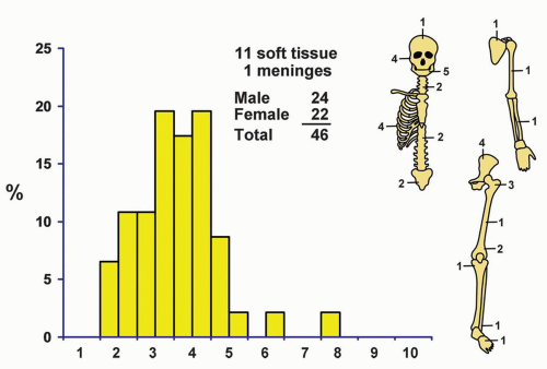 Figure 7.1. Distribution of mesenchymal chondrosarcomas according to age and sex of the patient and site of the lesion. |
RADIOGRAPHIC FEATURES
Radiographs show a lytic destructive process. Although a few of the lesions are purely lytic, most contain mineral. Many lesions have the radiographic features of conventional chondrosarcoma: intramedullary tumor with calcification, expansion of the bone, and cortical thickening. The tumors generally have poor margination, which suggests a malignant tumor. Mesenchymal chondrosarcomas that occur in the soft tissues typically contain stippled areas of calcification (Figs. 7.2 & 7.3).
The radiographic features of mesenchymal chondrosarcoma are not specific; however, they usually suggest a malignant tumor, probably of cartilaginous derivation.
GROSS PATHOLOGIC FEATURES
The tumor is typically gray-to-pink, firm or soft, and usually well defined. In rare instances, it is lobulated. The tumors vary in size, up to 14 cm in greatest dimension, and most contain hard, mineralized material that varies in amount from scattered foci to prominent zones. Some are cartilaginous, at least in part. Zones of necrosis and hemorrhage may be seen. Tumors of the soft tissue tend to be sharply circumscribed. Part of the lesion may appear soft and fleshy, similar to most sarcomas. However, most of them contain calcific foci (Fig. 7.4).
HISTOPATHOLOGIC FEATURES
Related posts:

Stay updated, free articles. Join our Telegram channel

Full access? Get Clinical Tree


