Radiographic evaluation—Hamada classification
Hamada I
3
Hamada II
12
Hamada III
22
Hamada IV
12
Hamada V
3
Supraspinatus
Ruptured
56
Infraspinatus
Totally ruptured
31
Partially ruptured
25
Goutallier classification (if not ruptured)
Goutallier I–II
0
Goutallier III
10
Goutallier IV
15
Teres minor
Atrophied
28
Absent
17
Normal
11
Hypertrophic
0
Statistical Analysis
Based on the relatively small sample size and no prior basis to assume the variables analyzed form a normal distribution, we used nonparametric tests to assess whether there were any statistically significant differences. To evaluate differences between preoperative and postoperative variables, a Wilcoxon signed-rank test was used, whereas a Wilcoxon rank-sum test was used to determine if any pre-, intra-, or postoperative variables correlated with outcomes. Medians and interquartile ranges were used to describe the central tendency and dispersion for the variables under study. Statistical significance was set at the 0.05.
Procedural Rationale
The rationale behind this procedure has been outlined in detail in our previous work [1–4]. The natural horizontal balance of the shoulder involves four internal rotator muscles (latissimus dorsi, teres major, pectoralis major, and subscapularis) and only two external rotator muscles (infraspinatus and teres minor). As a superior rotator cuff tear extends posteriorly, the integrity of the external rotators is compromised. In this situation, the key muscle maintaining horizontal shoulder balance is the teres minor. When this muscle becomes torn, atrophied, or fatty infiltrated, there is no other muscle available to compensate for active external rotation.
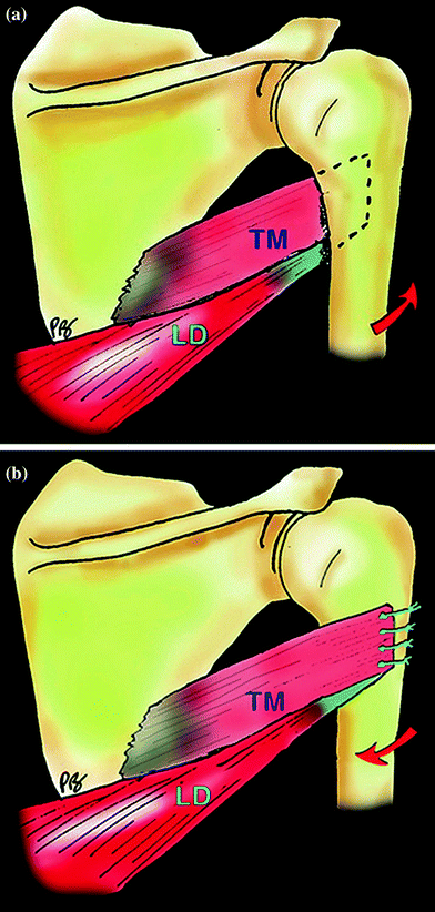
An RSA medializes and lowers the humerus, placing the deltoid at maximal mechanical advantage to compensate for the deficient rotator cuff. While this results in restoration of active anterior elevation and abduction (vertical muscle balance), it is ineffective in restoring active external rotation (horizontal muscle balance) when both the infraspinatus and teres minor are functionally absent. In previous series, the presence of atrophy or fatty infiltration of the teres minor has proven to be a negative prognostic factor for outcomes of RSA in cuff tear arthropathy patients [12–15]. Performing an LD/TM transfer restores the horizontal balance of the shoulder by transforming two of the internal rotators into external rotators (Fig. 26.1). In other words, the ratio of internal versus external rotator muscles is improved from 4 versus 0 to 2 versus 2. Combining the two procedures (RSA + LD/TM transfer) simultaneously restores both planes of motion (Fig. 26.2).


Fig. 26.1
Schematic demonstrating the principals of the modified L’Episcopo transfer. The latissimus dorsi (LD) and teres major (TM) are changed from internal rotators of the humerus (a) to external rotators of the humerus (b) by adjusting their point of insertion. This replaces the external rotation function lost by the deficient infraspinatus and teres minor

Fig. 26.2
Schematic demonstrating the combined effect of the reverse shoulder arthroplasty with latissimus dorsi/teres major transfer. Note the restoration of vertical balance (forward elevation, abduction) provided by the deltoid and restoration of horizontal balance (external rotation) provided by the tendon transfer
Surgical Technique
All procedures were performed by the senior author (PB) or under his direction through a deltopectoral approach in the beach-chair position. The incision is extended distally from that of a standard RSA, allowing release of the upper half of the pectoralis major tendon. All patients with an intact biceps tendon at the time of surgery also underwent a biceps tenodesis. The latissimus dorsi and teres major tendons are identified posterior to the pectoralis and released from the humerus. Care is taken to identify and protect the radial and axillary nerves during mobilization of the transferred tendons. Four heavy non-absorbable sutures are used to capture the latissimus and teres major tendons to facilitate transfer and fixation. A “tunnel” is created through blunt dissection posterior to the humerus to allow for tendon passage. Our preference is to prepare the transfer prior to performing the RSA. Once the final RSA is implanted, the transferred tendons are secured to the humerus. The patient is then immobilized in a position of 30° abduction and 30° external rotation for six weeks.
Our early practice was to transfer the two tendons to the anterolateral aspect of the humerus, bringing the two tendons to the lateral border of the bicipital groove to maximize the possible excursion that could result in external rotation. However, we had several patients who complained of an overtensioning effect that significantly limited internal rotation and prevented them from performing basic daily functions, such as manipulating the zipper on their pants. In some female patients, we also noted that the two tendons were weak (particularly the teres major, which has very little tendon at its humeral insertion) and difficult to reattach to the bone. These concerns led us to modify our technique through the years by: (1) harvesting the two tendons with a periosteal flap to reinforce the tendon-to-bone repair, (2) securing the transferred tendons through transosseous drill holes to provide the additional security of a bone bridge, and (3) transferring the combined tendons to the lateral margin of the humerus, (i.e., a more posterolateral location) to decrease the restriction on internal rotation.
Results
Complications and Reoperations
There were nine complications in 9 patients (16 %). One patient suffered a postoperative contralateral C7 palsy attributed to intra-operative positioning, which resolved with conservative treatment. Four patients experienced a humeral fracture under the prosthesis, 3 of which were treated with fracture fixation and maintenance of the prosthesis. Two patients developed prosthetic instability, one of whom had an RSA at another center before we performed his transfer. One patient suffered from a deep Propionibacterium acnes infection requiring a complete prosthetic exchange but without compromise of the transfer. The last patient was presented with persistent pain and stiffness that required revision consisting of arthroscopic AC joint resection, lysis of adhesions, and revision biceps tenodesis. The revision rate was 11 % (6/56). Two patients objectively ruptured their tendon transfer, one after a fracture and one after shoulder dislocation.
Radiographic Evaluation
Fourteen patients (26 %) demonstrated evidence of scapular notching, with seven patients rated as grade 1, three patients rated as grade 2, and four patients rated as grade 3, according to the Nerot–Sirveaux classification [15]. Twenty-six patients (47 %) demonstrated evidence of scapular spur formation.
Clinical Evaluation
Clinical follow-up averaged 44 months (range: 12–111). Two patients demonstrated a persistent deficit in external rotation and were considered failures of the tendon transfer. Because our goals were to evaluate the results of a combined procedure, these patients were excluded from functional statistical analysis.
A comparison of preoperative and postoperative outcomes can be found in Table 26.2. This demonstrates a statistically significant improvement in all measures, both objective and subjective. At final follow-up, 49 of 54 patients (91 %) were either satisfied or very satisfied with their surgical result.
Table 26.2
Comparison of preoperative and postoperative mean values
Preoperative Mean | Postoperative Mean | Mean delta | p-Value | |
|---|---|---|---|---|
AAE | 94° | 147° | 53° | <0.0001 |
AER1 | −18° | 10° | 28° | <0.0001 |
AIR | 5 | 3.8 | −1.2 | <0.0001 |
VAS | 5.5 | 1.1 | −4.4 | <0.0001 |
SSV | 29 % | 72 % | 43 | <0.0001 |
ADLER | 9 | 25 | 16 | <0.0001 |
Constant | 32 | 64
Stay updated, free articles. Join our Telegram channel
Full access? Get Clinical Tree
 Get Clinical Tree app for offline access

|


