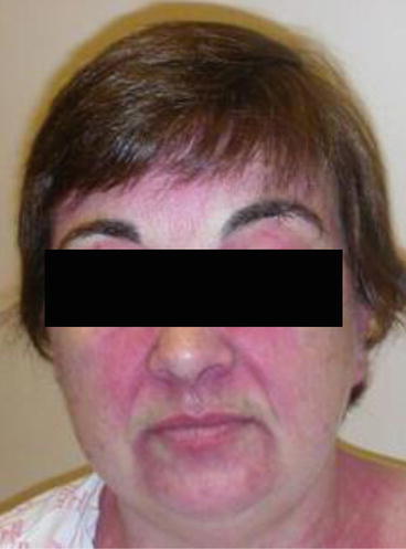
Fig. 5.1
Malar erythema from rosacea with characteristic papules and pustules
Lupus erythematosus: The malar rash is classically associated with acute cutaneous lupus erythematosus (ACLE) or subacute cutaneous lupus erythematosus (SCLE). The malar rash of ACLE does not tend to show very much epidermal change (other than perhaps minimal scaling), thus, the presence of crusting or erosions is more indicative of other disorders. This rash typically involves the nasal bridge but spares the nasolabial folds (Fig. 5.2). As mentioned, there are no pustules, although papules can occasionally be found. These patients can also have involvement of other photodistributed areas, such as the chest and arms. Typically, these patients are experiencing a flare of their SLE and thus have other LE-related symptoms. The malar erythema of SCLE is associated with more scale, and usually there is associated involvement of the chest, back, or arms. One can typically find more classical annular or psoriasiform configured lesions, and they are characteristically associated with hypopigmentation. The erythema of SCLE has a telangiectatic quality not typically seen in other causes of malar erythema.
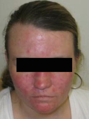
Fig. 5.2
Malar erythema from acute LE demonstrates confluent erythema with edema and sparing of the nasolabial folds
Dermatomyositis (DM): Patients with DM can have a malar erythema that is indistinguishable from LE-related erythema. Clues for DM include involvement of the eyelids, edema, predilection to involve the nasolabial folds (Fig. 5.3), associated involvement of other areas (especially the scalp), and a dysesthetic burning and itch. The rash of DM also has a characteristic violaceous color.