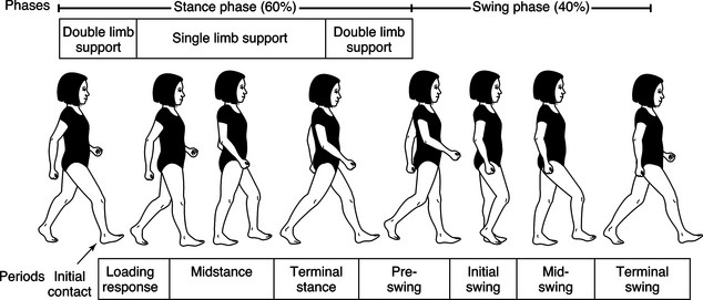
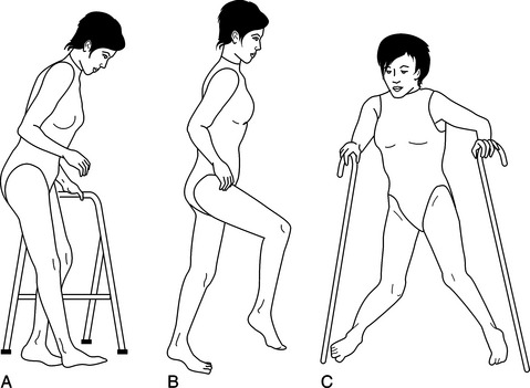
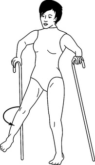
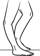
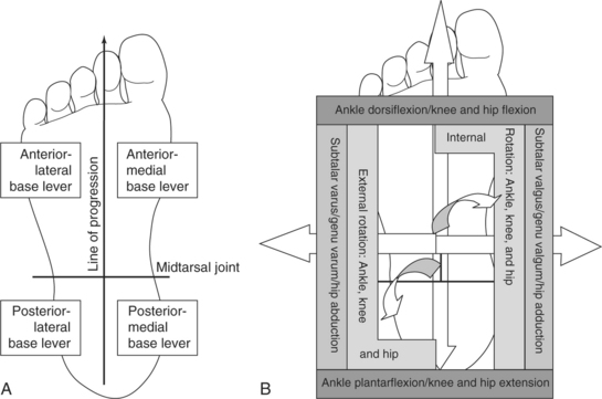
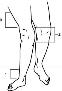
Lower Extremity Orthotics

CHAPTER 17


Lower Extremity Orthotics


Role of the Occupational Therapist


Historically, occupational therapy has primarily been involved in the provision of upper extremity orthotic services. However, with increased emphasis on the multidisciplinary team approach the scope of occupational therapy practice has expanded to include lower extremity orthotic care as it impacts the acquisition of occupational performance. It is important to delineate the role of occupational therapy in lower extremity, as opposed to upper extremity, orthotic treatment programs. In upper extremity orthotic practice, occupational therapists typically design, fabricate, fit, and supervise functional training. In effect, occupational therapists provide seamless delivery of orthotic services. Occupational therapists manage every stage of the upper extremity orthotic delivery process and are therefore able to adapt each step to individual needs and specific functional requirements.


In contrast, occupational therapists are not direct providers of lower extremity orthotic care. Typically, orthotists design, fabricate, and fit lower extremity orthotic devices and physical therapists provide functional gait training with lower extremity orthoses. Occupational therapy collaborates in the delivery of lower extremity orthotic services to ensure that the orthosis is designed to facilitate occupational performance at each stage of development.


Any given lower extremity device may address a biomechanical goal such as providing a stable base of support. The device may also address a functional gait training goal such as decreasing knee hyperextension during stance. However, if the orthosis does not also address the occupational performance goal (such as donning and doffing the device independently) the person may discard the orthosis. It is the occupational therapist’s role to anticipate such performance issues and initiate effective intervention before design and fabrication decisions have been completed. Occupational therapy clinicians are clearly the strongest advocate of advancing occupational performance goals.


Biomechanical and gait considerations are irrelevant when the lower extremity orthosis is rejected because it is too difficult to apply, interferes with activities of daily living, or is designed without consideration of individual strengths and weaknesses. With pediatrics, lower extremity orthoses can impact occupational performance differently and may be focused on the development of balance and equilibrium as a foundation for skill development and motor milestone acquisition. A lower extremity orthosis to position the hips may provide a stable base of support and help the child sit for longer periods of time to facilitate independent eating or writing skills. The same orthosis may not provide sufficient mobility for crawling and transitional movements unless it is designed to address these functional skills as well.


This chapter does not describe specific criteria used to design and fabricate orthoses, as do other chapters in this text. As the orthotist exclusively manages the fabrication process in lower extremity orthotics, it is not necessary or appropriate to cover this topic. However, if the occupational therapist is to be involved as part of the clinical team making decisions regarding lower extremity orthotics he or she must have a working knowledge of basic principles and terminology used by both physical therapists and orthotists in the decision-making process. This chapter is designed to provide a basic understanding and is not intended to be definitive or comprehensive in nature. Those with an interest in developing further expertise in this area should invest in additional educational seminars and reference texts.


Definition and Historical Perspective


An orthosis is generally identified as any orthopedic device that improves the function or structure of the body. Evidence of orthotic applications has been found as early as 2750 BCE [Bunch and Keagy 1976]. Excavation sites have uncovered mummies with various splints still intact. Historically, increased focus and advancements in orthotic design have centered on civil strife and periods of war. Today, continued advancements in the ever-expanding world of technology promote the development of new orthotic designs, materials, and components.


Purpose and Basic Function


The four basic functions of an orthosis include support and alignment, prevention or correction of deformity, substitution for or assistance of function, and reduction of pain. An orthosis is designed to address any one or all of these basic functions. Additional indications for lower extremity orthotic intervention are listed inBox 17-1.


General Applications


In the lower extremity, orthoses are prescribed for a variety of reasons and may be used on a temporary or permanent basis. For example, knee orthoses are often prescribed postoperatively to restrict joint motion while healing of soft-tissue structures occurs. After the rehabilitation phase, the knee orthoses are then either discontinued or used only during periods of increased physical activity.


In contrast, the young child with a diagnosis of spina bifida may require extensive orthotic designs to enable him or her to stand and walk. Such systems will be adjusted and replaced with growth but will play an ongoing role in maximizing and maintaining the child’s ambulation skills. In both instances, orthoses play a critical role in maintaining the structural integrity of the anatomy and the functional ability of the person. The relationship between structure and function is discussed in detail later in the chapter.


Basic Biomechanical Principles


Individual Evaluation


Prescription criteria for lower limb orthoses are based on a thorough pre-orthotic evaluation of the person. Components of this evaluation process are listed inBox 17-2. Observational gait assessment (OGA) is performed with the person wearing shorts and a snug T-shirt. This allows the observer to relate the function of the lower limbs to the stability of the upper torso. Walking trials are performed with the person barefoot and then with any existing orthosis or ambulation aids being used. Biomechanics laboratories can offer a detailed analysis to assist in evaluation of the person’s gait deviations with the use of force plates and high-speed cameras. However, these facilities are sparse and the cost of such a procedure is often prohibitive. Individual goals, motivation, and available social support contribute to the success of any orthotic program.


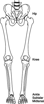
Walking Requirements


Walking is a complex series of muscular interactions that manipulate the skeletal structures in specific sequential and reciprocal patterns. As a result, normal walking occurs in a mechanically effective and energy-efficient manner (Figure 17-1). The gait cycle consists of heel-strike to ipsilateral heel-strike of the same limb, covering periods of double and single limb support as well as swing. The gait cycle can be further divided into eight specific phases: initial contact, loading response, mid-stance, terminal stance, pre-swing, initial swing, mid-swing, and terminal swing [Perry 1992]. Critical events occur at each phase of gait to enhance stability and encourage functional mobility. Extensive texts are available on this topic, which is not the focus of this chapter. Therefore, a brief summary is presented in the following as it relates to lower extremity orthotic treatment programs.


Normal Gait


Limb stability is one of two basic components of walking. Each lower limb, in turn, must be effectively aligned and controlled to accept the transfer of body weight, support and balance the entire body mass independently, and then transfer the weight to the contralateral limb as it comes in contact with the ground. Sixty percent of the gait cycle is spent in periods of single and double limb support, when one and then both lower limbs support the body weight. Limb advancement is the second basic component serving to help translate the body mass forward and is dependent on the support provided by the contralateral stance limb. Swing phase accounts for 40% of the gait cycle. These actions occur reciprocally while maintaining forward momentum of the body through space with minimal metabolic costs.


Although most lower limb joint motions occur in the sagittal plane (i.e., hip and knee flexion and extension, ankle dorsiflexion and plantar flexion), motions also occur in the coronal and transverse planes. This serves to streamline the gait cycle and limit the translation of the center of mass (COM) to minimal vertical and horizontal excursions. As a result, a decrease in the displacement of the top-heavy trunk segment is evidenced and energy is conserved. The most efficient gait pattern for each individual occurs at a self-selected walking speed, when the person is trying neither to increase nor decrease normal walking speed.


Pathologic Gait


A pathologic gait pattern often develops secondary to neuromuscular deficits, joint instabilities, pain, disease processes, congenital involvements, and many other conditions (Figure 17-2). Excessive or insufficient joint motions occur and lead to exaggerated or inhibited movements of the body throughout the gait cycle. The normal walking speed of the individual will also be affected. Increases in energy costs tend to promote decreased walking speeds and further increases in energy costs.


Clinical training in observational gait assessment ensures the identification of all pathologic gait deviations in need of orthotic intervention. Commonly observed deviations include drop-foot, hyperextension or genu (knee) recurvatum, lateral trunk lean, anterior knee instability, genu varum or genu valgum, pelvic instability, increased lordosis, tone-induced equinovarus, pes planus, and so on. Observational gait assessment also evaluates many factors, such as overall symmetry, step lengths, loading patterns, width of the base of support, weight transfer, terminal stance stability, swing phase function, and trunk alignment, just to name a few. Other pathologic factors include joint contractures, muscle weakness, and disturbed motor control programs.


Functional Motions (Compensations)


Most persons acquire a pattern of walking that serves a functional rather than an efficient purpose, when compared with normal parameters. For example, when dorsiflexion capabilities at the ankle are lost, a person experiences a drop-foot condition. The toes drag on the ground during swing phase and contact first with the ground during the loading phase. Persons will adopt a gait pattern that provides ground clearance for the plantar-flexed foot during swing phase. This is accomplished by excessive hip and knee flexion (i.e., steppage gait) or by excessive hip circumduction (Figure 17-3). Both of these compensations achieve a functional result; specifically, ground clearance during swing phase. Although these compensations occur without conscious thought and may allow for ambulation, the need for an orthosis is supported by increased energy costs and safety concerns.


Dysfunctional Motions (Detractors)


Although such clinically observable gait deviations detract from effective and efficient gait, some dysfunctional motions are more notably difficult to compensate for in a functional manner. Hyperextension (or “back knee”) is such a condition. This occurs as the knee moves in the posterior direction during mid-stance, often secondary to weakness of the quadriceps and/or posterior calf muscle groups. Without hyperextension, the person experiences uncontrolled knee flexion and collapse.


As a side note, a plantar-flexion contracture is often noted upon examination as either the primary cause or result of the posterior knee moment and serves to limit tibial advancement over the base of support during stance phase. Genu recurvatum (Figure 17-4) is the long-standing result of hyperextension, when permanent damage to the posterior soft-tissue structures of the knee has occurred. This gait disruption affects the efficiency of gait because the lower extremity is forced posteriorly as the body mass is attempting to move anteriorly over the limb. Working at odds, large truncal deviations are noted and energy costs increase dramatically.


Orthotic Design Principles


General Concepts


Principles of lower extremity orthotic design consider the interaction of anatomic and mechanical structures. The mid-tarsal, subtalar, talocrural, knee, and hip joints are evaluated in regard to the individual joint alignment and range of motion (ROM), as well as the interaction of these five major joints during the task of walking. Application of mechanical joints can serve to encourage, restrict, or eliminate potential joint motion.


Considerations of joint position, relief of pain, restriction of motion, or relief of weight bearing relate to the effect of the application of forces. Force is applied to effect an angular change at a specific joint in one or more of the three planes of motion. As a result, care is taken to apply forces over pressure-tolerant areas (i.e., soft tissue or adequate surface areas), with relief of force over pressure-sensitive areas (i.e., bony prominences). Mechanical leverage is another important concept, as specific force is increased with shorter lever arms and decreased with longer lever arms. Properly applied forces over adequate surface area with long lever arms exert less force for effective joint control than do orthotic designs with short mechanical lever arms.


Additional orthotic considerations include weight, cost, adjustability, ease of application, cosmesis (appearance), maintenance, and the necessary training required to ensure successful outcomes [Bunch et al. 1985, Kottke and Lehmann 1990]. These factors should be discussed thoroughly by the rehabilitation team and the individual before a final orthotic design is determined.


Joint Alignment


All five major joints (mid-tarsal, subtalar, talocrural, knee, and hip) of the lower extremities exhibit triplanar joint motion (Figure 17-5). Still, they work together during walking to allow progression of the body in the desired direction. These motions occur largely through the sagittal plane, specifically hip and knee flexion with ankle dorsiflexion and plantar flexion. Alignment of skeletal joint levers is a critical component of efficient and effective ambulation.


Mechanically speaking, the foot can be divided into anterior and posterior portions, with the mid-tarsal joint serving as the connection (Figure 17-6). Through mechanical leverage, the long anterior lever is used to control ankle dorsiflexion, knee flexion, and hip flexion. The short posterior lever limits ankle plantar flexion, knee extension, and hip extension. Medially, the first metatarsal ray and the medial heel act as medial levers to support proximal joint structures and prevent subtalar valgus and genu valgum, limit hip adduction, and discourage excessive internal rotation of the entire limb. The fifth metatarsal ray and the lateral heel serve as lateral levers to prevent subtalar varus and genu varum, limit hip abduction, and discourage excessive external rotation of the entire limb.


Joint Stability


The base levers of the foot are similar in concept to the foundation of a house. A square and true foundation always provides the best support. An unstable base of support has the potential to create larger moments of instability at proximal joint structures. For example, a pronated (flat) foot has clinically observable features: hind-foot valgus, mid-foot collapse, and forefoot abduction. Mechanical assessment reveals a loss of the anterior-medial forefoot and posterior-medial hind-foot base levers. Therefore, the ankle joint is subjected to excessive dorsiflexion, internal rotation, and medial displacement; the knee is subjected to flexion, internal rotation, and valgum moments; and the hip joint is subjected to flexion, internal rotation, and adduction moments. An effective orthotic design that returns joint stability and proper alignment to the mid-tarsal and subtalar joints will serve to effect positive alignment changes at the ankle, knee, and hip joints.


Three-point Force Systems


As mentioned earlier, the application of force is used to effect angular changes at deviated joints. A three-point force system (Figure 17-7) effects an alignment change with two forces working in opposition to a counterforce (or fulcrum). The counterforce is positioned on the convex side of the joint deviation, close to the joint requiring an angular change. The opposite two forces are positioned proximal and distal to the counterforce, on the side of the joint concavity. The greater the linear distance between opposing forces the less pressure is required to maintain the angular correction.


Components and Materials


A wide variety of mechanical joints and materials are now used in orthotic designs. Mechanical joints are designed to mimic anatomic joint function, and care is taken to approximate anatomic joint alignments. Orthotic joints are manufactured in a variety of metals and plastics, with unique design features to control available ROM. Special strapping is often used to enhance alignment and control. Material selection is based on the desired design characteristics of the orthosis. Material properties range from extremely flexible to extremely rigid. Different types of foam padding are available in variable thickness, density, and durability to meet different mechanical design requirements. Keep in mind that an orthosis is a mechanical device that requires periodic checkup and maintenance to ensure proper mechanical functioning and to extend the longevity of the orthosis.


There are a number of questions that need to be answered before selecting the appropriate material. Does the material need to be flexible or rigid? Lightweight or heavy duty? Inexpensive? Temporary or permanent? What is the length of time required for preparation? Can the materials be easily cleaned? Can the material be maintained easily? Can the material allow for easy donning and doffing? Although all concerns are usually not met at the first orthotic fitting, much frustration on the part of the team and the client can be avoided if all are involved early in the process and share the same end-product outcome goals.


Material selection is based on the desired design characteristics of the orthosis. Material properties range from flexible to rigid, lightweight to heavy-duty, inexpensive to costly, limited to prolonged durability, and minimal to extensive fabrication preparation. Each of these factors must meet the client’s anticipated activity level as well as demands of aesthetics, ease of cleaning, maintenance, and don/doff procedures. These various, at times conflicting, considerations compound the difficulty of clinical decision making in orthotic selection. Appropriate choices lead to acceptance and independence, whereas unwise selection may cause not only rejection of a device but unintended injury to the client.


The process of material selection weighs each of these factors in consideration of the client outcome or goal. The selection process can be relatively apparent. If the desired outcome is to gradually increase active motion of a postoperative knee joint over several weeks, the clinician selects an orthosis that is lightweight, inexpensive, adjustable, of limited durability, and easily fabricated. If, however, the desired outcome is to protect the same knee joint from ACL tear reoccurrence during a contact sport such as soccer the material selection process focuses on an orthosis that is highly durable, of rigid construction (probably involving extensive/costly fabrication), very low profile in design to minimize interference during the activity, easily cleanable to reduce skin irritation, and cosmetically appealing (as the orthosis would be highly visible).


More challenging material selection processes involve competition between factors. The client may desire a lightweight, flexible, inexpensive, easily fabricated device to reduce foot drop. These material selections, however, will not address the underlying problems of a severe osteoarthritic ankle, which may necessitate selections the client finds unacceptable. At times, material selection focuses on a single factor that supercedes all other considerations. To reduce hypertrophic scarring that accompanies severe burns, it is essential that a continuous hard smooth surface be applied 24/7 to achieve the clinical outcome of scar prevention/reduction.


All other factors of comfort, aesthetics, ease of don/doff procedures, fabrication procedures, and costs are subservient to the factor of material texture in the determination of the clinical outcome. For some conditions, hand function is severely limited. No matter how effective any knee/ankle/foot orthosis (KAFO) functions are in improving gait, if the client is unable to don/doff the device efficiently and consistently it will be rejected as not practical for their daily routine. It is imperative that therapists communicate their concerns during the material selection process, during the orthotic evaluation.


Orthotic Classifications


Three main types of orthoses are available. Prefabricated (or over-the-counter) designs are manufactured in a variety of sizes and offer immediate application, reduced cost, and simplicity. They are generally used as evaluative tools or for temporary use because the fit/function and the durability of these products are limited. Custom-fitted designs require a more involved measurement and fitting procedures to obtain better fit and function. Used for moderate involvements, custom-fitted designs are prescribed on both a temporary and definitive basis. Custom orthotic designs are the most sophisticated, requiring extensive measurements, castings, fitting, and follow-up procedures from a skilled orthotist. Most often prescribed on a permanent (or definitive) basis, a custom orthotic design is made to specifically fit the individual and is therefore more expensive to manufacture. The result, however, is a more intimately fitting device with greater joint control, limb stability, and improved function and mobility.


Another aspect to consider is whether a static or dynamic splint is warranted. The most common uses of static splints are (1) to support joints that need to be immobilized, (2) to help prevent further deformity, and (3) to prevent soft-tissue contractures. As an immobilizing force, these rigid devices place and maintain a joint or joints in one position while allowing healing of a fracture or an inflammatory condition. It is optimal to place and hold joints in a position of function. Static splints prevent further deformity by maintaining a controlled stretch on the affected joint.


Conversely, dynamic splints allow motion of a joint within a prescribed range while supporting other joints. Primary uses of dynamic splints include (1) to act as a substitute for lost motor function, (2) to correct a deformity, (3) to provide controlled motion, and (4) to facilitate fracture alignment and wound healing. The dynamic action comes from hinges or springs placed in line with the joint to be acted upon. The tension of the hinge/spring can be set at a prescribed ROM and tension can be based on the use and desired outcome. Dynamic splints are more complex in their design and fabrication than static splints. Understanding the functions and uses of the static and dynamic splint will lead to the most effective choice.


Orthotic Terminology


The terminology for lower extremity orthoses is based on the anatomic area affected by the orthosis. An arch support or shoe insert is called a foot orthosis (FO). An orthosis designed to address drop-foot is called an ankle/foot orthosis (AFO). The orthoses may be further defined by descriptive terms such as rigid FO or thermoplastic AFO, or may be designated by the function performed such as ground reaction AFO or stance-control KAFO. Consistency in terminology ensures effective communication within the rehabilitation team. Standard abbreviations for lower limb orthoses are listed inTable 17-1.


Duration of Use


Therapists and orthotists both make devices intended to support, align, stretch, control, and replace the function of compromised joints and muscle groups. Often the decision of whether the device is made by a therapist or orthotist is based on the amount of time the device is expected to be used. Splints that are usually made by therapists are fabricated from low-temperature splinting materials with a life span of 3 to 6 months. These splints can be technically demanding but the duration of time in the orthosis is limited. Low-temperature thermoplastic splinting materials do not require a scan or cast of the body part to be fabricated, and are contoured directly over the client’s skin or stockinette-covered skin.


Lower extremity splints, fabricated using components that are easily assembled, may also have metal attachments. They are assembled using hand tools normally available in the splinting lab. Splints used for smaller body parts such as the arms, hands, neck, and sometimes the lower leg can be changed fairly easily as the client moves through the rehabilitation process. Some rehabilitation centers use low-temperature materials to fabricate full body splints for postoperative positioning. However, working with large sheets of plastic that easily stick together is quite cumbersome and technically demanding.


Orthotists, on the other hand, use high-temperature thermoplastic material that is heated to 325° F or more. These devices (usually referred to as orthoses) are ordered when the orthosis is expected to be worn for more than 6 months or to withstand greater forces as in weight bearing. High-temperature thermoplastic material is too hot to be contoured directly over the client’s skin, and thus a scan or cast is taken of the affected body part to acquire a detailed mold. The mold is closed and filled with plaster. The high-temperature thermoplastic is then heated and draped over the mold under a vacuum to create the orthosis. After cooling, trim lines are drawn, and the plastic is removed using a cast cutter.


Edges are finished and buffed using grinders and routers, and any metal hinges or hardware are applied before the straps are attached. The high-temperature plastic and metal components are made to last for years, and can withstand the forces of spasticity and muscle weakness for longer periods of time than low-temperature splinting materials. A variety of high-temperature thermoplastic choices is available and these are chosen based on needed characteristics. Lower extremity orthoses have to withstand the repeated forces of weight bearing over long periods of time. Spinal orthoses are often made of more flexible high-temperature thermoplastic materials with full foam liners. Orthotic designs intended for high activity over long periods of time are best fabricated by an orthotist from high-temperature materials.

Stay updated, free articles. Join our Telegram channel

Mar 13, 2017 | Posted by in PHYSICAL MEDICINE & REHABILITATION | Comments Off on Lower Extremity Orthotics

Full access? Get Clinical Tree

Get Clinical Tree app for offline access