Fig. 42.1
(a, b) Radiographs of a liposarcoma of the femur. Uncharacteristic lucent mass in the medullary compartment with some endosteal scalloping and undefined limits
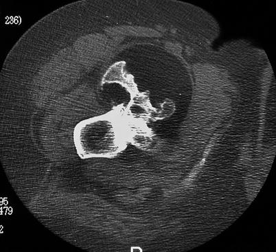
Fig. 42.2
CT scan of surface liposarcoma of the proximal femur. Well-circumscribed lucent lesion distorting the host bone external shape
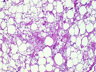
Fig. 42.3
Low-power microscopic view of well-differentiated liposarcoma
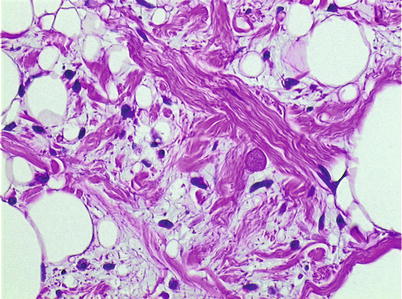
Fig. 42.4
Medium-power microscopic view of well-differentiated liposarcoma. Atypical cells are more easily found in the spindle cells septae of the lesion
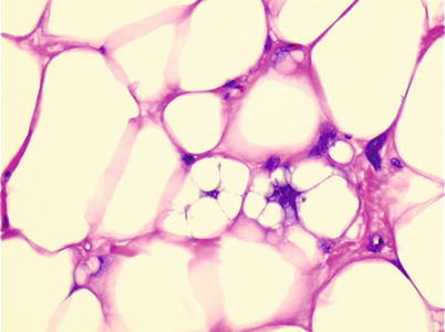
Fig. 42.5
High-power microscopic view of well-differentiated liposarcoma. There may be only a few atypical cell nuclei. Multivacuolated adipocytes and lipoblasts are also seen
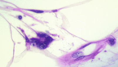
Fig. 42.6
High-power microscopic view of well-differentiated liposarcoma. Multiple atypical nuclei in adipocyte
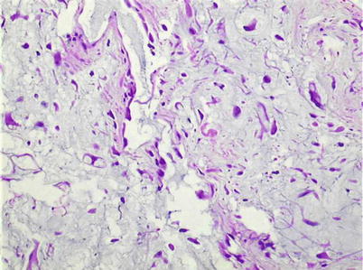
Fig. 42.7


Medium-power microscopic view of pleomorphic liposarcoma
Related posts:


Stay updated, free articles. Join our Telegram channel
Full access? Get Clinical Tree
