, Paola Catalano1, Andrea Piccioli2, 3, Maria Silvia Spinelli4, 3 and Federica Zavaroni1
(1)
Anthropological Service, Soprintendenza Speciale per il Colosseo, il Museo Nazionale Romano e l’Area Archeologica di Roma, Rome, Italy
(2)
Oncologic Center “Palazzo Baleani” Azienda Policlinico “Umberto I”, Rome, Italy
(3)
The Italian Society of Orthopaedics and Traumatology (SIOT), Rome, Italy
(4)
Department of Orthopaedic and Traumatology, Catholic University Hospital, Rome, Italy
4.1 Introduction
Carla Caldarini5 , Paola Catalano5, Andrea Piccioli6, 7, Maria Silvia Spinelli8, 7 and Federica Zavaroni5
(5)
Anthropological Service, Soprintendenza Speciale per il Colosseo, il Museo Nazionale Romano e l’Area Archeologica di Roma, Rome, Italy
(6)
Oncologic Center “Palazzo Baleani” Azienda Policlinico “Umberto I”, Rome, Italy
(7)
The Italian Society of Orthopaedics and Traumatology (SIOT), Rome, Italy
(8)
Department of Orthopaedic and Traumatology, Catholic University Hospital, Rome, Italy
4.1.1 Evolution of Orthopaedic Surgery in the Treatment of Degenerative Conditions
During the twentieth century, the development of orthopaedic surgery led to a more solid definition of the orthopaedist’s boundaries and competences. It started at the beginning of the 1900 from Alessandro Codivilla’s intuition, and his stating that the orthopaedist had to deal with the diseases of the musculoskeletal system [1]. However, the main pathologies involving orthopaedists in the first few years of last century were the congenital and childhood ones, those resulting from poliomyelitis, and the bone complications of tuberculosis. Later on, the treatment of degenerative diseases focused mainly on the treatment of coxoarthritis, among which the worth mentioning are consequences of congenital dysplasias, and knee arthritis. Up until the 1960s, arthritis surgery mainly consisted of osteotomies, with the aim of modifying the axes and the load areas of the joint cartilage, and arthrodeses, which inevitably led to poor functional results in large joints, even though it acted directly upon the cause of the arthritic pain. In 1962, Sir John Charnley described his hip prosthesis, thus changing the approach to degenerative surgery for ever [2]. In parallel, only after several attempts, John Insall improved the knee prosthesis with a condylar design in the 1970s [3]. Interestingly, in about a decade the approach to the treatment of coxoarthritis changed completely in the records of the national congress of the Italian Society of Orthopaedics and Traumatology (SIOT). In the 49th Congress in 1964 on the surgical treatment of coxoarthritis, osteotomies, arthrodeses, and many interventions on the soft parts were the pillars of the orthopaedist’s cultural background, while more modern therapies such as acetabuloplasties and arthroplasties were only beginning to develop. In 1969, many orthopaedists already reported on their experience in prosthetic implants, and the scientific community was split between “conservatives and prosthesisizers”. Since then, the development of more and more sophisticated prosthetic implants has characterized orthopaedic surgery, which nowadays faces the issue of revision surgery and complication management [4]. Another innovation in the treatment of degenerative conditions has certainly been the implementation of arthroscopic techniques of the large joints, which has been widely considered as a technological innovation of the last few years, even though a 1912 contribution from Severin Nordentoft has just been found: at the 41st congress of the German Surgery Society in Berlin he presented his contribution on laparoscopy, cystoscopy, and knee endoscopy, followed after a few years by Takagi (from Japan), and Bircher (from Switzerland), who mainly concentrated on joint endoscopy [5]. Finally, the last few years have seen a growing interest in new treatment approaches towards degenerative arthritis, through the implementation of bioengineering in surgery, in particular with studies on osteocartilage transplants, mesenchymal cells, biomaterials, and growth factors.
History of Medicine: Arthritis Care
Valentina Gazzaniga and Silvia Marinozzi
When there is no damage or evidence of trauma or tumour, joint pain is generally explained as a sign of weakness induced by other diseases, or ageing, or inappropriate life styles, and leads to the accumulation of cold and corrupt matter in the peripheral parts of the body. In arthrosis and arthritis, of pathological origin, or deriving from wear, load or stress, the procedure is the same as in case of inflammation. According to the Hippocratic theories pathological processes are a sign of imbalance among the fundamental elements of the body, fluids, qualities, and solid parts, which compromises the health of the whole body. Without any apostemas, swelling, or localized clotting, the corrupt humours are drained, in particular the cold ones (such as phlegm and black bile), with drainage and calefaction treatments to re-establish normal body heat and bring the body back to balance. The therapy involves enemas, emetic medications, cupping, diets and localized treatments, such as warm compresses, and in the most complex cases, where the pain is very sharp, and there is motor difficulty and a chronic condition, the cauterization of the areas where there is visible or hypothetic clotting of pus (Celsus, De re medica, lib. IV, 33–35;). Galen mainly relates about chronic pain and inflammation of the femoral joint. There is a nosological overlap between low back pain and skeletal pathologies, so everything is attributed to an inflammatory process involving the whole limb due to plethoric processes to be treated with warm compresses, baths, rest, appropriate exercise, and drainage treatments, both pharmacological and surgical, which is phlebotomy. The disease is not caused by the pituous humour per se, but by the fact that it stays raw: the treatment varies according to the patient’s temper, age and physical strength. First palliative medications, which do not cool or warm too much, to ease the pain, then bloodletting from the popliteus muscle or from the heel. According to Galen, acrid medications, largely widespread in his time, have to be avoided, because they cause excessive dryness, which would solidify the dense pituous humours, that undissolved continue to cause illness; he prescribes palliative medications and composite oilcloths, providing several recipes for oilcloths and mixtures, mainly based on turpentine, tar, sulphur, and “spices” such as cardamom, colocynthis, ruta, and other “warming” substances (Galen, De Compositione medicamentorum, X,2-3 – K.XIII: 331–341; XIV: 756).
4.2 Femoro-acetabular Impingement
4.2.1 Morphological and Paleopathological Description of the Subject
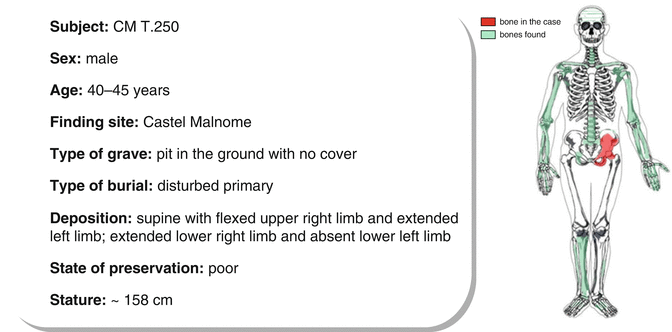
The skeleton pertaining to a mature male, it is in a poor state of preservation: only a few fragments of the skull are preserved and regarding the inferior appendicular skeleton, only the proximal epiphysis of the left femur, a portion of the left fibula, the right tibia and fibula, and part of the feet bones are left. The examination of the dental disease shows a big abscessual cavity at the alveoli of the maxillary right lateral incisor and canine. The right first premolar and contra-lateral canine and second premolar are affected by destructive decays; moreover, many maxillary and mandibular teeth were lost intra-vitam. On the clavicles, at the origin of the deltoid and at the insertion of the pectoralis major muscle, enthesopathic modifications, of osteophytic type, due to repeated movements of the shoulder, are bilaterally detected. Strong insertions at the origin of the brachii muscle are observed on the humeri, especially on the right, because of a repeated flexion of the forearm on the arm. In reference to the lower limbs, the right tibia is medio-laterally flattened (platycnemia); the flattening is probably due to a strong and prolonged exertion of the calf muscle. The only remaining small diaphyseal portion of the left fibula shows a severe periostitis with scabrous surface and exuberant reactive bone production. The shoulder joints show some mild arthritic degenerations: on the acromial and sternal facets of the clavicles and on the humeri heads, a mild porosity is spread, with poor marginal ostheophytes. The enthesopathy examined at the insertion of the supraspinatus muscle of the right humerus, would confirm a frequent rotation of the humerus on the scapula. The column shows poor lipping on the ventral margins of the vertebral bodies; a posteriorly expelled hernia is also detected on the inferior endplate of a thoracic vertebra.
4.2.1.1 Description of the Lesion
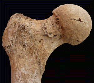
The present case shows a deformity of the head-neck compartment of the left femur now known as “cam” type (Figs. 4.1 and 4.2) that suggests femoro-acetabular impingement.
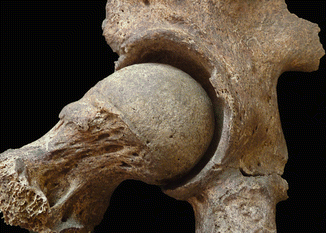
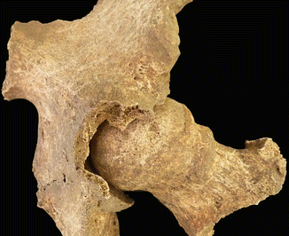

Fig. 4.1
Femoro-acetabular impingement posterior view

Fig. 4.2
Femoro-acetabular impingement anterior view
Femoro-acetabular impingement (FAI) is a condition that is more and more frequently treated and detected nowadays in adult active patients complaining of hip pain. The Medical Subject Headings thesaurus of the US National Library defines FAI as a “condition where a pathological mechanical process causes hip pain when morphological anomalies of the acetabulum and/or the femur, combined with vigorous hip motion (above all at the extreme degrees), lead to repeated collisions that damage the soft tissue structure within the joint itself”.
The essential features of this definition are as follows:
Morphological anomalies of the femur or acetabulum
Anomalous contact between the two structures
Supra-physiological movements leading to this contact
Recurrence of these movements and the mechanical insult
Damage of the soft tissue
FAI can be further subdivided into two categories, depending on whether the deformity concerns the acetabulum or the femur, called respectively “pincer” and “cam deformity”, or both. The typical clinical picture is a progressive hip pain in a young active patient, which can irradiate laterally to the trochanteric region, medially to the adductor muscles region and, more rarely, to the gluteal region or to the knee. In the past some authors [6] hypothesized that only 10 % of hip arthritis could be accounted as purely primary and that in more than 90 % of cases the pathogenesis was due to other causes (mechanical, inflammatory, metabolic, biological). Ganz et al. [7] stated that more than 90 % of hip arthritis was due to mechanical pre-existing conditions such as an acetabular dysplasia and FAI.
4.3 Primary Arthritis
4.3.1 Primary Arthritis of the Hip in Coxa Vara
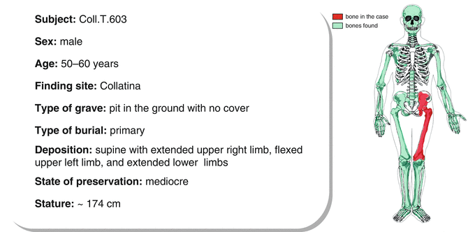
4.3.1.1 Morphological and Palaeopathological Description of the Subject
The subject, averagely built, has humeral euribrachy and strong platymeric femurs. The skull is elongated (dolichocranic) and flat (chamaecranic); the frontal crests are very diverging, the orbits are wide (hypsiconchic), and the nose is long and narrow (leptorrhine). The porosity of the outer table of the skull (cribra cranii) and of the roof of the orbits (cribra orbitalia), together with hypoplastic lines on the enamel of some mandibular teeth, suggests stress episodes that may have impaired the normal development of the subject. The maxillary bones show alveolar resorption processes due to the loss of all dental components (edentulism) intra-vitam; the central incisors and the lateral left one of the mandibular bone were also lost ante mortem, and an abscess can be observed on the buccal side of the left mandibular second molar. The carious lesions on the remaining teeth are widespread, but moderately severe, with the exception of a destructive decay detected on the right second incisor. The limited presence of alterations in the muscle-ligament insertions and the moderate degree of degenerative phenomena observed on the whole skeleton, suggest that the subject was not involved in hard work. The only enthesopathy observed on the upper limbs regards the insertion of the brachii muscle of the ulnas; this muscle is used for flexion of the forearm on the arm. Even though the rachis is incomplete, the fusion of two thoracic low vertebrae can be observed. The trochanteric fossae of the femurs, on the insertion surface of the obturator externus muscle, are affected by exostoses; the gluteous insertions are marked, bilaterally. An extension of the articular surface of the head of the right femur, which can be seen on the anterior surface of the neck (Poirier’s facet) (Fig. 4.3) [8], may be the result of a contact between the caput femoris and the margin of the acetabular fossa during flexion and extension movements of the limb [9]. A further etiological factor can be the acute and repeated muscular stress of the iliopsoas muscle, which presses on the medial margin of the cervical eminence. The anterior surface of the patellae is affected by ossifications of the quadriceps tendon, mainly on the right side. The diaphyses of the lower limbs, in particular of the tibiae and fibulae, show osseous remodeling due to acute or chronic inflammatory processes, and this affects the cortical bone, as well. The surfaces are irregular and rugged, due to the deposition of a new bone in the form of striae and plaques. The anterior crest of the left tibia is particularly well developed, forming a protruding plica (Fig. 4.4), compared with the 4.6 mm cortical bone underneath.

