, Paola Catalano1, Valentina Gazzaniga2, Silvia Marinozzi3 and Federica Zavaroni1
(1)
Anthropological Service, Soprintendenza Speciale per il Colosseo, il Museo Nazionale Romano e l’Area Archeologica di Roma, Rome, Italy
(2)
Department of Sciences and Medico Surgical Biotechnologies, History of Medicine Unit, Sapienza-Università di Roma, Rome, Italy
(3)
Department of Molecular Medicine, History of Medicine Unit, Sapienza-Università di Roma, Rome, Italy
1.1 Funerary Archaeology and Field Anthropology
Carla Caldarini4 , Paola Catalano4 and Federica Zavaroni4
(4)
Anthropological Service, Soprintendenza Speciale per il Colosseo, il Museo Nazionale Romano e l’Area Archeologica di Roma, Rome, Italy
Bio-archaeological studies and historical documents are a great tool to reconstruct the lifestyle and health conditions of the ancient populations, and to understand the correlation between man and the environment over the course of time. The Anthropological Service has taken part in the environmental protection activity of the Soprintendenza Speciale per il Colosseo, il Museo Nazionale Romano e l’Area Archeologica di Roma. This has contributed to outline the biological history of Roman society, in particular that of the Imperial age. In the last decades, new excavation methods applied to the human skeletal remains have helped to collect valuable information on Roman sepulchres, especially those found in the Suburb, because of the large number of civil buildings built after the urban development. These data, together with those deriving from in-depth laboratory investigation, are helping to understand the complex biological landscape of the ancient Roman population with its bio demographic and social processes.
What did the Suburb look like during the Imperial age? As Rodolfo Lanciani [1] wrote: “In its best days, Rome was practically one with the nearby towns of Veii, Nomentum, Tivoli, Palestrina, Tusculum and Bovillae. Villas, vineyards, rural estates with country houses shaped both small and large areas creating a wide populated park around Rome”.
In recent years, the investigation on a large number of broad excavations held by the Soprintendenza, has been throwing light on wide areas whose countless archaeological contribution had been ignored [2]. The anthropological material, firstly collected during the systematic excavation of the necropolis [3] and then through laboratory investigation [4] is a considerable help to understand how the funeral services were held and how life was in the Urbs and in its surroundings, in an attempt to analyse funeral customs and everyday lifestyle through a chronological and socio-historical perspective which has been overlooked for a long time [5].
Moreover, an inter-disciplinary analysis allows to establish a connection between the everyday life conditions, the diseases, and the specific therapies of the communities that the anthropological finds belonged to. This book is an example of the valuable contribution given by medical historians and pathologists to this aim.
The anthropological analysis of a burial must begin “in the field”, at the time when it is found (Fig. 1.1). Taphonomy is the branch of knowledge which studies the processes (chemical, physical and biotic) that the body experiences between the individual’s death and the body’s discovery, based on the reconstruction of the action and interaction of the different factors which have interfered with it, from the moment of the deposition until its finding.
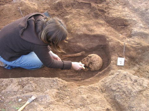
Fig. 1.1
Burial during excavation
The information obtained through the taphonomic study of skeleton, reported case by case on an appropriate database, need a careful examination of the position of every skeletal element and the record of the condition of every joint. This is how the deposition can be defined as primary or secondary. In a primary deposition, the body has been buried, immediately after its death, in its ultimate place, where it decayed; at the time of unearthing, the skeleton must be in its original burial position, accepting for changes of position due to the action of taphonomic agents or secondary handling. Further changes of positions after the burial can be detected, whether designed, as in profanation, or accidental, linked to natural causes, as either the passage of animals or water damage [6]. A secondary deposition refers to a burial performed at two or more different times: the finding of the skeleton, and so the definitive deposition, happens in a different place from the one of the decay. The bones may have moved from their earlier position simply due to gravity, which pulls the bones down during the decomposition of the corpse, because of the absence of the soft tissues and ligaments which normally hold them together.
These movements can therefore be interpreted in order to reconstruct the original position of the body, thus highlighting the funerary practices of deposition: for instance, we will be able to determine the presence of perishable funeral structures (as cushions, coffins or dressings), or whether the deposition occurred in the ground, and so the decomposition took place in a restricted space, or in a sarcophagus, with the decomposition in a hollow space.
During the excavation process, experts should not only collect taphonomic information, but also determine, when possible, the gender and age at death of the subject, detect any pathological alterations and perform some measurements, because the frequent frailty of the finds can cause a serious loss of information, if the findings are recorded after the exhumation. This is the reason why most of the methods which will be briefly described below are usually applied in the field first and then in a laboratory in a more thorough and detailed way [7].
1.1.1 Determination of Sex
The parts of the skeleton showing features with the highest degree of sexual dimorphism are the skull (Fig. 1.2a, b) and the pelvis (Fig. 1.3a, b); in particular, the latter is structured in women to cope with pregnancy and birth giving. Because a subject rarely shows all the typical features of its gender, we have considered a combination of features, taking into account their importance in the diagnosis, as described by various authors [8–11]. For some subjects, a gender diagnosis is sometimes impossible, because the findings are fragmented and incomplete.
Fig. 1.2
(a) Female skull. (b) Male skull
Fig. 1.3
(a) Female coxal bone. (b) Male coxal bone
1.1.2 Estimation of Age at Death
Various methods are used to calculate the age of death in adults, considering individual variability. The skeleton of an adult does not suffer great changes over time, so the age cannot be determined through the analysis of a single criterion; therefore, it is estimated applying several different methods and balancing the various findings, thus obtaining a time range in which the age of the subject under study is likely to be found.
One of the most noteworthy methods commonly used is the study of the degree of tooth wear, because the occlusal surface of the teeth gets thinner with age (Fig. 1.4a, b). Other factors determine tooth wear: the diet, the presence of dental-alveolus pathologies and some jobs involving the use of teeth [12].
Fig. 1.4
(a) Mild dental wear. (b) Severe dental wear
The degree of welding of the ectocranial sutures has also been detected, based on the progressive fusion of the cranial sutures, until a complete obliteration, generally occurring in older age (Fig. 1.5a, b). Amongst all, Meindl and Lovejoy’s method was used [13]. Then the auricular surface of the ileum was examined: in young subjects, the auricular surface is characterized by wavelet and diagonal rifts and the bone tissue is granular; with ageing, the diagonal rifts and the scratches decrease, the grainy quality of the bone tissue is gradually replaced by small areas of dense and compact tissue and by porous portions, the margins becoming irregular and torn. In old age, the surface shows porous and eroded areas, the margins are irregular and in the retro-auricular area strong bone protrusions and osteophytes can be observed. Amongst all we used Lovejoy et al.’s method [14].
Fig. 1.5
(a) Non-ossified ectocranial sutures. (b) Ossified ectocranial sutures
Moreover, the morphology of the pubic symphysis has been detected – in young subjects it is wrinkled, while with ageing it becomes irregular, with porosities, erosions and ossifications. We used Burns’ method [15]. Then, the sternal end of the ribs has been examined – under 15 years of age the surface is flat and mildly wavy; with ageing it alters until it assumes a V shape, while the margins rise. After 55 years of age, the degenerative process prevails, until the margins fray and the porosity increases. Iscan et al. [16] studied these phenomena, but the method is difficult to apply due to the extreme frailty of the ribs.
The diagnosis of sub-adult’s age at death (less than 20-year-old dead subjects) has been determined by the examination of the degree of development and dental eruption: this method identifies the different development phases and the dental eruption, an almost constant feature up to 14–16 years of age. We have used Ubelaker’s chronology of dental eruption [17]. In addition, we have detected the size of the diaphyses of the long bones, and compared the values obtained with some charts based on the tight connection between the length of the long bones (measured without epiphyses) and the subject’s age. However, it is important to bear in mind that measurements are always affected by the individual and population variability [18]. Considering the degree of ossification of the growth cartilage, we can estimate the subject’s age if it is less than 24 years: in most cases, at this age the diaphysis and epiphysis of the bones, which compose the skeleton, are completely fused. In this study we used the standards given by Krogman e Iscan [19], France e Horn [20], Suchey et al. [21], Ubelaker [18], Iscan [22].
1.1.3 Osteometry
All the bones which were intact enough to be measured were analysed osteometrically in a laboratory, in order to evaluate the length, width, section and circumference. We followed Martin and Saller’s measurement procedure [23]. For every measurement we have indicated Martin’s number and the commonly used abbreviation. The measurements are expressed in millimetres (mm) and taken with appropriate anthropometric instruments, such as the osteometric board (Fig. 1.6), the round curved compass to measure width and length, the digital calibre to measure sections and the millimetre strip to measure the circumference.
Fig. 1.6
Femur measurement with an osteometric board
We used the osteometric dimensions to elaborate the height [24, 25] and the main indices of strength and section of the long bones, to try to highlight morphometric differences due to dynamic and environmental factors, such as stress from physical exercise or nutritional deficiency.
The main indices are as follows:
1.1.3.1 Cranial Indices
Cephalic index (horizontal) (8/1 × 100): ratio between width and maximum length. This determines the elongated (dolichocranium) or short (brachycranium) shape of the skull.
Height index (vertical-longitudinal) (17/1 × 100): ratio between the height measured at the bregma and the maximum length. This determines the degree of flatness of the cranial vault.
Frontal transverse index (9/10 × 100): ratio between the minimum and maximum frontal diameter. This determines more or less the diverging shape of the superior forehead.
Fronto-parietal index (9/8 × 100): ratio between the minimum and maximum frontal width.
Gnathic index (alveolar) (40/5 × 100): ratio between the length of the face and the nasion-basion length. This determines the degree of face prognathism.
Orbital index (52/51 × 100): ratio between the height and length of the orbit. This determines more or less a circular shape of the orbit.
Nasal index (54/55 × 100): ratio between width and height of the nose. This determines a longer or shorter shape of the piriform aperture (long and narrow: leptorrhine; short and large: camerrhine or platyrrhine).
1.1.3.2 Post-cranial Indices
Strength index of the clavicle (6/1 × 100): ratio between the circumference measured in the mid-diaphysis and maximum length.
Humerus diaphysic index (6/5 × 100): ratio between the minimum and maximum diameter measured in the mid-diaphysis. When the value of the index is close to 100 the two diameters are equal and the diaphysis is round shaped (euribrachy): when the minimum diameter is considerably smaller than the maximum, we have a flatness of the humerus (platibrachya), maybe caused by intense workload of the biceps and deltoid muscle.
Humerus strength index (7/1 × 100): ratio between minimum circumference and maximum length.
Radius diaphyseal index (5/4 × 100): ratio between the sagittal and transverse diameter measured in mid-diaphysis. The smaller the values, the more protruding the interosseus crest due to greater pronation and supination movements of the forearm.
Ulna olenyc index (13/14 × 100): ratio between the superior transversal and the superior dorso-volar diameters; this shows the transversal flatness in the superior section of the ulna.
Ulna diaphyseal index (11/12 × 100): ratio between the dorso-volar and the transversal diameters; the smaller the values, the more protruding the interosseous crest.
Ulna strength index (3/1 × 100): ratio between minimum circumference and maximum length.
Femur pillar index (6/7 × 100): ratio between the antero-posterior and transverse diameters, measured at mid- diaphysis. A high index means a strong development of the linea aspera (pilaster), often linked to thigh muscular work.
Femur platimetric index (10/9 × 100): ratio between the antero-posterior and transverse diameters measured below the lesser trochanter. An index lower than 85 means the antero-posterior crushing of the diaphysis superior third (platimery) generally linked to a strong development of the trochanters due to a great biomechanical stress.
Femur strength index ((6 + 7)/2 × 100): ratio between the sum of the antero-posterior and transverse diameters and the physiological length. A value higher than 12.5 means a good strength of the lower limb.
Tibia diaphyseal index (9/8 × 100): ratio between the sagittal and transversal diameters measured at mid- diaphysis.
Tibia knemic index (9a/8a × 100): ratio between the sagittal and transversal diameter measured at the nourishing passage. A value lower than 65 indicates the diaphysis flattening in a medio-lateral direction (platicnemia); the crushing is generally due to the work of the calf muscles, maybe due to a prolonged and intense use of the legs.
1.1.4 Dento-alveolar Diseases
The anthropological study has also covered the state of the dento-alveolar complex. The study of the oral pathologies is really important in anthropology because of the great amount of information that can be obtained; from the conditions of the teeth, we can obtain data on the eating and hygiene habits of the subjects. Any presence of tartar, dental decay, abscesses, parodontopathies and intra-vitam losses has been recorded.
Tartar, made of limestone deposits due to plaque mineralization, has been quantified in three degrees: small, average and large amount.
Dental decay easily develops when saliva is slightly acidic. Organic acids derive from carbohydrate fermentation due to bacteria in the plaque. If acidity is high, especially in a sugar-rich diet, it can break down the tooth mineral, and lead to enamel and dentin perforation. Dental decay is classified depending on location (occlusal, buccal, lingual, mesial, distal), severity (superficial, affecting the dentin, perforation or affecting the crown) and affected tooth area (crown, neck, root).
An abscess is caused by an inflammation reaching the tooth canal through decay, wear and tear or fracture. A tooth abscess can be detected through a drainage canal at the root apex. Also in this case we have recorded presence and position (buccal, lingual, apical).
Parodontopathies are caused by inflammatory processes involving the tissues surrounding and supporting the tooth; they cause a progressive decrease of the alveolar margin, exposing the root and allowing for bacterial penetration. The alveolar re-absorption can occur evenly and with the same extension in most teeth, or it can be localized around a single tooth. Moreover, a dental pocket can occur at the neck or at root level, inclined to an apical extension and leading to an abscess as a final outcome [26]. The alveolar re-absorption is classified as light when the junction-cement and alveolar margin is below 4 mm and as severe when the distance is greater.
Finally, there is an alveolar re-absorption and a bone remodelling where the tooth has been lost (intra-vitam loss).
1.1.5 Non-specific Stress Index
Many factors affecting metabolism, such as diseases, acute or chronic nutritional deficiencies, due to either a lack of consumption (direct) or of absorption (indirect), lead to an alteration of the bone tissue diagnosed through a macroscopic examination. The anthropological study has also detected these alterations (non-metrical indices on functional stress and on disease).
Porotic hyperostosis is a macroscopic porosity seen on the orbital roof (cribra orbitalia) and/or on the external surface of the cranial vault (cribra cranii). This alteration is related to anaemic states of different aetiology. For instance, hereditary haemolytic anaemias, anaemias following infectious or parasitic diseases, or anaemias due to an iron deficiency [27].
Exostosis is a bone formation in the external hearing canal and can appear as an irregular bone mass. Several clinical studies [28] have shown the onset of exostoses with a prolonged water exposition. Some types of dermatitis, traumas, haemorrhages and senile changes can lead to external otitis and so to exostoses.
Auricular osteophytosis, is due to light inflammation of the external ear. It manifests itself as a slight bone proliferation around the external acoustic meatus, leading to the formation of an irregular edge with small-size osteophytes. Both diseases, when present, have been indicated with a degree of severity: mild for up to 2 mm formations, severe for more than 2 mm formations.
Enamel hypoplasia manifests itself as lines or little wells on the teeth surface, and it is caused by the interruption or slowing down of the enamel formation during the amelogenesis, that is the growth phase of the permanent teeth (the period from birth to 6–7 years of age). These alterations are caused by non-specific stress factors (such as malnutrition or diseases), so they are useful indicators of the health condition and quality of life of a population [29].
Periostitis is an inflammation of the periosteum. The pathology can be mainly observed on the tibia surface, due either to microtraumas or venous stasis [30].
1.1.6 Musculoskeletal Markers, Arthropaties, Traumas
The bone tissue remodelling due to intense physical exercise or hard work is defined through ergonomic markers or MSM (musculoskeletal stress markers) [31] and MOS (markers of occupational stress). Among them, we can find enthesopaties, arthropaties, non-metrical stress and trauma markers.
Entheses (from the Greek enthesis, “introduction”) are areas of muscular and ligament insertion [32]; in the absence of a specific biomechanical stress, they manifest on the bone as imprints (wrinkles, protrusions, cracks), and they represent variable indicators of strength. Under stress, the same points of insertion can show osseous proliferations and/or erosions, defined as osteophytosis and osteolysis. These alterations are due to a non-specific pathological state of the enthesis, generally called enthesopaty, which can be present or absent, while the strength signs are always recognizable.
These modifications can have a mechanical, metabolic or inflammatory origin.
Osteophytosis and osteolysis are also due to age or idiopathic factors of the bone’s response to different stimuli: the border between a healthy and a pathological form is difficult to define.
Arthritis is a degenerative pathology of the joints of the post-cranial skeleton and of the intervertebral discs of the rachis, occurring with a marginal opening (lipping), osteophytosis, porosity and eburnation. These alterations are the result of a discrepancy between requested work and work capacity of the joint. Arthritis location, age of onset, spread and development have a multifactorial origin; even though the causes can have a systemic nature (ageing, hereditary factors, sex, obesity, etc.) and/or a local one (overload and joint dynamics alterations), biomechanical stress is the necessary condition for the development of arthritis.
As arthritis gets worse, and leads to a direct contact between the bone surfaces, these can become shiny (eburnation) and show stripes parallel to the direction of the joint movement, often accompanied by porosities [33].
Other types of non-metric markers of functional stress, defined as “discreet” MOS (Markers of Occupational Stress) [34] include all the extensions of the joint surfaces (squatting and Poirier optional facet, sacroiliac and Allen fossae), the lack of fusion of the ossification centres (acromial bone) and the vastus medialis muscle notch (also called Messeri patella), due to overload.
A Poirier’s facet [35] is an extension of the articular surface of the head of the femur on the anterior surface on the neck, maybe due to a contact between the caput femoris and the margin of the acetabulum cavity, when the limb flexes and extends. A further etiologic factor can be a hard and repeated stress of the iliopsoas muscle, which presses on the medial margin of the cerebral prominence.
Both a prolonged crouched posture and a repeated flexion of the foot, when marching on rough ground [36], can cause the development of optional facets (squatting facets) at the level of the tibio-talar joint [7]. These features occur as an interruption of the continuity of the anterior margin of the tibial distal epiphysis.
The optional sacroiliac facets are round areas, localized at the level of the first or second sacral foramen, sometimes having their equivalent on the iliac tuberosities. These modifications are generally connected to loads carried on the back, with an increase of lumbar lordosis [7].
The Allen fossa, situated on the anterior face of the femoral neck, is connected to extensions and flexions of the limb [31]; it is the outcome of the contact of the rectus muscle tendon on the anterior part of the femoral neck. Its presence is due to an upright posture [33], associated to ambulation on steep ground [37–39].
Bipartite acromion, the potential outcome of an over use and load of the rotator cuff [40], is the lack of fusion of the acromion with the scapular spine.
Messeri patella [41, 42] is the result of the modification of the kneecap morphology. A frequent flexion of the legs or a habitual crouched position involves the quadriceps muscle tendon; its over stress leads to a lesion of the superior–lateral surface of the basis.
When studying the rachis, Schmörl hernias (the extroflexion of the inner fleshy nucleus from the vertebral body) have been taken into account. These nodes are related to traumas, ponderal overloads during adolescence or pre-existing conditions (infections, rickets and osteoporosis). These lesions are mainly found on the central portion of the lumbar vertebrae plateau and on the lower thorax [43]. The spinal column has also shown some osteophytoses, fractures and fusion of the bodies.
Among the most easily detectable alterations in the anthropological samples, traumatic lesions provide information on the environment and on the characteristics and hardness of the work carried out. The environment can cause particular physical traumas, recognizable on the skeleton: for instance, a high frequency of both wrist and ankle fractures can be due to working on irregular ground.
The bone remains have shown some traumatic lesions, such as fractures, dislocations, haematomas or pulled muscles. These have been recorded on an appropriate database, separating fractures/dislocations from tendon/muscle traumas.
Traumas are classified into accidental (due to life style), deliberate (due to violence), ritual, punishment (amputation) and therapeutic (surgical); their frequency, localization and severity can provide “traumatological patterns”, useful for the analysis of the socio-cultural features of the ancient populations. Fractures are complete or partial interruptions of the bone structure integrity, leading to a healing process seen in new formations (osseous callus).
A dislocation is a permanent relocation of the joint surfaces. It is called complete when the loss of connection between the two surfaces is absolute. When partial contact remains, it is called incomplete or sub dislocation. Its aetiology can have a traumatic origin if occurring after a violent trauma which moves the ends of the bones; innate if due to a malformation of the ends of the bones; degenerative (pathological) if due to capsular ligament lesions.
1.1.7 Skeletal Markers of Working Activity
These markers increase in number and intensity with age, but also respond to mechanical stress [44]. The data regarding these alterations can be a useful parameter to trace a population’s lifestyle [45, 46], thus contributing to the identification of load patterns and work division based on gender [47–49], or social status.
The muscular–skeletal stress markers have been recorded considering Hawkey [50] and Mariotti [51, 52] with three grades of development for every enthesis. The first grade (1), characterized by a weak and average development, is divided into three sub types: light (1a), low (1b) and average development (1c). The second (2) corresponds to a marked manifestation and the third (3) to a really strong development of the tract. These grades are all physiological: absent or normal modification.
Enthesopathies, when present, are divided in proliferative osteophytic forms (OF) (Figs. 1.7 and 1.8) and erosive, osteolytic forms (OL) (Fig. 1.9). The suggested pattern for their classification includes three grades that define the pathological stages. The first corresponds to light porosities and exostoses smaller than 1 mm; the second one to several areas of erosion and exostoses between one and four mm in size; the third one to marked exostoses, bigger than four mm. When there is no enthesis, or in case of a bad preservation status, the examined skeletal section has been recorded as “ND” (that stands for:. not detected). For the discrete markers, the presence/absence of traumas, fractures and arthropathies has been detected; for fractures and arthropathies also the site has been evaluated, recording the cases of politraumas.
Fig. 1.7
Enthesophytes at the insertion of the ulna triceps brachii. (Grave 176 Castel Malnome)
Fig. 1.8
Enthesophytes at the origin of the tibial soleus muscle (Grave 146 Castel Malnome)
Fig. 1.9
Erosion at the insertion of the costo-clavicular ligament (Grave 60 Quarto Cappello del Prete)
1.2 History of Medicine in Rome
Valentina Gazzaniga5 and Silvia Marinozzi6
(5)
Department of Sciences and Medico Surgical Biotechnologies, History of Medicine Unit, Sapienza-Università di Roma, Rome, Italy
(6)
Department of Molecular Medicine, History of Medicine Unit, Sapienza-Università di Roma, Rome, Italy
Ancient Rome is, for the historian of medicine, an extremely rich reality due to the theoretical development, the available and highly intellectual, methodological and technical written sources, and the rich evidence provided by the archaeological findings and remains [53].
The chronological period, the anthropological and palaeopathological study dwells on in this book is one of the most representative of the whole ancient times: in Rome, a rich Italics heritage of herbal and therapeutic competence faces the medical knowledge acquired in Greece since the classical times, rising increasingly complex pharmacological theories, whose peak is Galen of Pergamon’s “grades theory” but also the flourishing of encyclopaedic essays and manuals addressed to a wide and less refined public [55].
The worship of thaumaturgic gods, which only changes the name to the really wide number of Greek gods somehow involved in the treatment and in the promise of healing, is not only stifled by the importation of the rational medicine from Hippocrates, but, in a syncretistic way, it is enriched by the contributions from the Etruscan civilization first and then from the oriental esoteric religions; the survival of templar medical practices, which can be wholly superposable to the pre-classical and classical Greek ones, is well documented by Ovid’s poetical tale on the importation of the worship of Asclepius and, in more recent ages and in a geographical and cultural Greek background, by the rhetorician Aelius Aristides’ autobiographical Sacred tales, which tells his experience in healing.
As regards the rational medicine, the physicians claiming a medical training regularly gained in famous schools and teachers, have already learnt the lessons of the Hippocratic medicine suggesting a reworking which comes to an amazing complex degree (one of the most striking examples are Galen of Pergamon’s writings) [54].
The heritage of the Alexandrian medicine, whose history is halfway the third century B.C. in a very short-term and extremely important period, is seen in Rome with a meaningfully increased anatomical awareness compared to the Greek experience: the human body, which was conceptualized as a hollow vessel intended to hold fluids in almost all the Hippocratic writings, is seen as an elaborate system of parts interacting to each other in the innovative Galenic medicine, completely able to exploit the Alexandrian Herophilus’ and Erasistratus’ medical experience on dissection and autopsy. Every part of the body has got its own function, which leads to health if successfully achieved; the change in the anatomical structure involves an impairment and so a disease [55].
The medical sects question the method as well as the epistemological nature of medicine [56]; the Empire opens up to imported cultural phenomena and faces with experiences, the therapeutic ones too, from the conquered countries and from the ones with an active trade, leading to wonderful opportunities of theoretical and practical enrichment particularly seen in pharmacopoeial works, as the Compositiones, dated around 47–48 A.D. and written by Scribonius Largus, a physician operating under emperor Claudius. In these works new substances of vegetable, animal and, in small percentage, mineral origin, spread by the maritime trade, meet the earliest pharmacological expertise, raising a really varied pharmacopoeia, where features of folk medicine merge, as well as the scientific ones. [57] Moreover, in their preface letter sent to Callistus, Scribonius’ Compositiones are an example of how the Roman medicine achieves original results as regards the moral and ethic thought; the suggestion to judge every patient equal and worth of treatment, even the enemies of the homeland, enriches a moral “inner” tradition coming from the Hippocratic Oath with new elements [58].
So, a very rich “medical marketplace” [59], characterized by different professional competences and by a progressive refinement of the medical knowledge comparing to the Greek origin, practised together by physicians from the Hippocratic tradition, traditional healers, specialist surgeons, obstetricians, priests of Asclepius and Gods and heroes skilled in the art of medicine, military doctors and slaves.
Consulting the several available historical sources, Roman Imperial medicine looks like a field of knowledge deeply related to the civil and social life, a field where different expectations and waitings join with an increasingly rising market demand [60]. The geographical borders expansion with the high variability of the socio-economic status of the inhabitants of the Empire, also followed by the change of the risk factors, of the onset conditions and spread of infectious diseases, in a word of the whole pathological background (called “pathocenosis” by Mirko D. Grmek, who coined a highly important neologism for the future study of the ancient Greek–Roman diseases) makes the Roman world a privileged place to investigate on the sanitary ancient history.
This work has favoured some of the available sources, because of their connection to the history of the treatment of orthopaedic injuries and trauma: first of all, the Hippocratic treatises, which directly or indirectly regard the healing of fractured and dislocated limbs, head trauma and wound’s general treatment. As well-known, these works are not entirely attributable to the Kos master, although some of them were by the earlier reviewers, in particular Erotian, who lived under Nero, and author of a Lexicon, one of the first work trying to sort out the complex issue regarding the attribution of the works passed on under the name of Hippocrates. All these writings, however, share a rather early dating (end of the fifth – beginning of the fourth century A.D.) in unison with Hippocrates’ real life span and are the basis of the “technical” reflection on a Roman “specialized” orthopaedic competence. In particular, operating from the medical and encyclopaedic point of view, Galen and Celsus regard them the starting point to process again bone, skeleton and limbs technology and intervention.
Celsus (1st cent. A.D.), as well known, has not got a medical background, but he is a highly brilliant data collector showing the best example of technical refinement ever reached in the Roman encyclopaedic field in his work De Medicina; in his survived book on surgery many pages regard the orthopaedic techniques and the description of the tools used for the treatment of bone injuries in Rome during the first Empire [61].
Galen (130–200/210) is the well-known intellectual giant of medicine in antiquity; a versatile, very lucky and rich man, Marcus Aurelius’ doctor first and then his son Commodus’, he spends almost his whole life in Rome being on duty of the higher social classes. During his long Roman years, Galen collects books and makes up one of the most important medical library in the ancient times (destroyed by the Temple of Peace fire in 192 A.D. it was rebuilt by Galen himself, book by book, buying again what could be found on the market and writing “ex novo” his own works, when he didn’t manage to find a copy among friends and acquaintances). His work, which survived almost entirely through the Syriac and Arabic translations, includes several essays on the treatment of fracture and dislocation, the instruments and technique [62].
The available written sources are very rich. We can also add the support of the material history, thanks to archaeology [63]: the healing temples, consecrated to Asclepius and to a huge number of principal and secondary deities, all involved in disease healing, which carry on their task during the Empire, a task confirmed by the richness of anatomical ex voto (Fig. 1.10a–c), a wide field of documents on diseases and deformity affecting the Romans and the Empire inhabitants, although unable to originally repeat the typologies already documented by the Greek and Etruscan clay production in forms and genres [64]; the treatment centres for slaves and soldiers (above all established at the borders of the Empire, the valetudinarian are complex buildings where wounded soldiers are admitted and where a military competence is practised, also extended to the orthopaedic treatments) [65]. We can add some most singular events: the ruins of private surgeries, as the Surgeon’s House in Rimini, a second century A.D. building, where the Hellenic physician Eutyches lived and worked. When his house was destroyed by a fire, maybe in the third century after a barbarian raid, he left one of the richest surgical legacy of the ancient times; a hundred and fifty tools, of different material and dating, testifying their owner’s high specialized level, and, maybe, the “military” origin of his medical knowledge [66].
Fig. 1.10
(a) Roman ex-voto: hand and foot, ca. I-II. Cent. A.D. (Museo di Storia della Medicina, “Sapienza” Università di Roma). (b) Roman ex-voto: harm, ca. I-II. Cent. A.D. (Museo di Storia della Medicina, “Sapienza” Università di Roma). (c) Roman ex-voto: foot, ca. I-II. Cent. A.D. (Museo di Storia della Medicina, “Sapienza” Università di Roma)
Epigraphy helps to recreate the profile of the physicians working around the Empire, testifying the wandering features of the medical profession practised till the end of the Empire, excepting some cases concerning very famous physicians, settled for long periods in the same town. As regards, a slight difference between the practice of the Roman medical knowledge and its wandering organizational features in the Greek world, is the really wide dimension of the territories where doctors practice and the different cultures they meet: often coming from the eastern and Greek lands, not rare sepulchral statements testify their profession at the opposite edges of the great Empire.
Related posts:

Stay updated, free articles. Join our Telegram channel

Full access? Get Clinical Tree


