Fig. 15.1
An axial computed tomography scan of a type-C glenoid. Friedman’s line (a) is a line between the tip of the medial border of the scapula to the center of the glenoid joint, and the dashed line (b) is the axial diameter of the glenoid after reaming. The asterisk indicates the amount of bone reaming needed to provide a flat glenoid surface, and the dagger indicates the amount of bone left in the glenoid once the glenoid has been reamed flat

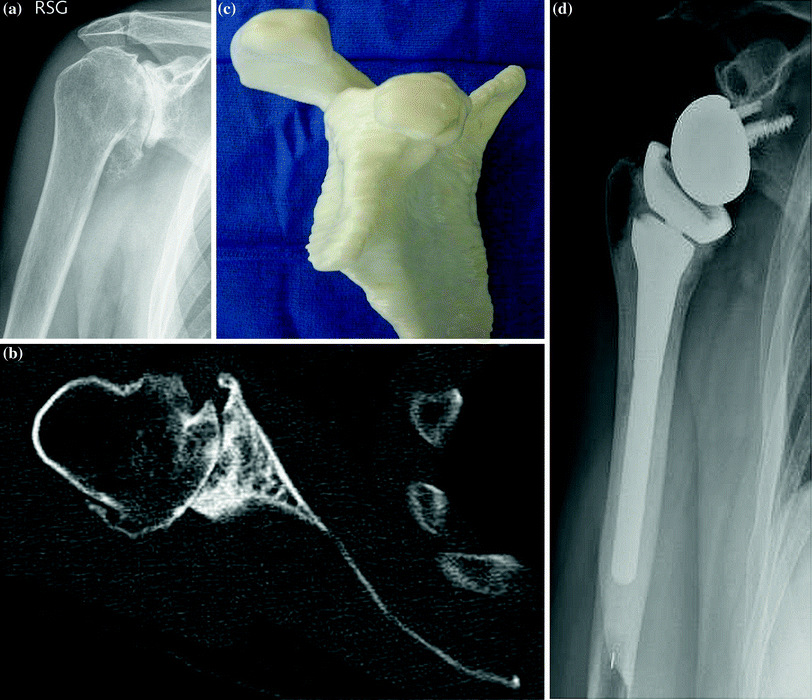
Fig. 15.2
Images of a patient with a type-C glenoid. a Preoperative anteroposterior radiograph. b Preoperative axial computed tomography scan. c Preoperative scapular model. d Postoperative anteroposterior radiograph obtained at 12 months after reverse total shoulder arthroplasty performed with reaming the glenoid flat with no bone grafting
Bone grafting with a reverse total shoulder prosthesis for patients with severe glenoid bone loss has also been reported to be successful in these patients [13]. Our preferred method of surgery for most patients more than 80 years of age who have degenerative arthritis of the shoulder without a rotator cuff tear is to implant a reverse prosthesis. Patients more than 70 years of age who have major rotator cuff thinning on MRI scanning also may be considered good candidates for a reverse prosthesis, especially if there is marked rotator cuff disease on MRI.
If an RTSA is performed in patients with an intact rotator cuff, there is little need to reattach the subscapularis tendon back to the lesser tuberosity. The incidence of instability in RTSA is the same whether the subscapularis tendon is reattached or not [22]. Edwards et al. [23] found that, with a Grammont type of prosthesis, instability was less when the subscapularis tendon was reattached than when it was not [23]. This finding might be prosthesis design-dependent because a prosthesis with a lateral offset and a higher head neck angle has a lower rate of instability than the Grammont prosthesis [24].
Key Points and Pearls
The use of a RTSA in patients with an intact rotator cuff has been reported to be successful in the presence of glenoid bone loss.
Although RTSA can be used in anticipation that a patient with rheumatoid arthritis might develop a rotator cuff tear, the complication rate in this population is high.
RTSA is indicated in patients with intact rotator cuff tendons but tuberosity malunions.
For an elderly patient with a thin rotator cuff on MRI or who is more than 80 years of age, consideration should be given to a RTSA once the patient is fully aware of the risks and benefits of this approach.
Younger patients with an intact rotator cuff and adequate glenoid bone stock should still have a traditional anatomical TSA.
References
1.
2.
Young AA, Walch G, Pape G, Gohlke F, Favard L. Secondary rotator cuff dysfunction following total shoulder arthroplasty for primary glenohumeral osteoarthritis: results of a multicenter study with more than five years of follow-up. J Bone Joint Surg Am. 2012;94(8):685–93.
Stay updated, free articles. Join our Telegram channel

Full access? Get Clinical Tree


