CHAPTER 34 Injury of the Flexors of the Elbow: Biceps Tendon Injury
INTRODUCTION
Except for epicondylitis, injury to the muscles or tendons about the elbow, as an isolated event, is rather uncommon.3,13,16,30,70 Distal biceps tendon injury, usually avulsion from the radial tuberosity, is the most common tendinous injury in this region, and the incidence seems to be increasing. Calcific tendinitis has been observed in the biceps tendon, but it is very uncommon.58
DISTAL BICEPS TENDON INJURY
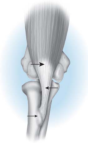
The biceps muscle-tendon complex may be injured at the musculotendinous junction by an incontinuity tear of the tendon and by a complete or partial tear or avulsion from the radial tuberosity (Fig. 34-1).
MUSCULOTENDINOUS JUNCTION
This is an uncommon injury and one rarely reported.68 The mechanism is similar for all biceps injuries—an eccentric load against a contracting biceps muscle. It may have a predilection for persons with encephalopathy, a condition present in many who experience triceps rupture.
AVULSION
By far, the most common injury is tendon avulsion, and complete avulsion is much more common than a partial injury. According to McReynolds,54 the first known diagnosis of a distal rupture was reported by Starks in 1843.
INCIDENCE
Avulsion of the biceps tendon at its distal insertion was reported in three of 100 patients with biceps tendon rupture who were studied by Gilcreest.25 The European literature suggests that distal avulsion injury accounts for approximately 3% to 10% of all biceps tendon ruptures.35 Only 24 cases were reported in a 43-year period after the original surgical descriptions by Johnson40 in 1897 and Acquaviva1 in 1898. Three hundred and fifty-five surgeons responded to a questionnaire Dobbie circulated in 1941, and only 51 cases were added.21 In addition, only three of this group had experience with as many as three cases. By 1956, the world literature contained 152 cases.27 At present, the injury is well known; either the incidence may be increasing or the lesion is recognized more often.*
More reports in the literature notwithstanding, we have encountered only two instances of involvement in a woman in the English literature.54 In addition, more than 80% of the reported cases have involved the right dominant upper extremity, usually in a well-developed man9,62 whose average age is about 50 years5,21,57 (range 2163 to 7021 years).
MECHANISM OF INJURY
In virtually every reported case,8,21,57 a single traumatic insult, often a force of 40 kg or more against resistance from an elbow in about 90 degrees of flexion, has been implicated. This mechanism, along with abuse of anabolic steroids, accounts for its surprisingly common occurrence in well-conditioned, healthy, but competitive weightlifters. Pre-existing degenerative changes in the tendon predispose to rupture.18,19 Acute pain in the antecubital fossa is noted immediately. Rarely, a patient complains of a second episode of acute pain several days later. Such a history suggests the possibility of an initial partial rupture or of secondary failure of the lacertus fibrosus.11,14 Occasionally, forearm pain has been reported, but it is considered rather uncommon.
ETIOLOGY
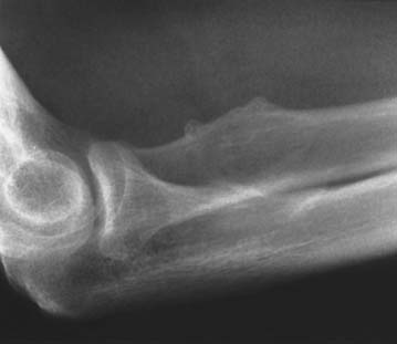
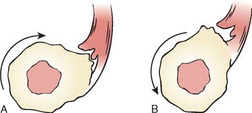
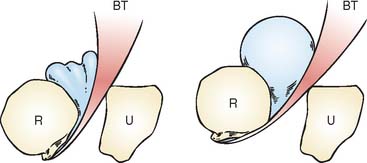
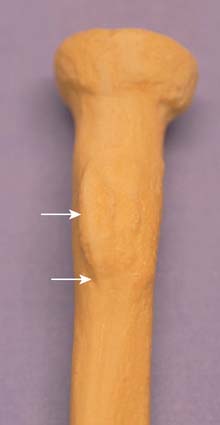
The cause of the injury has been discussed by several authors and is considered in detail by Davis and Yassine.18 The histologic pathology is that of a degenerative process, a finding consistent with the radiographic changes of spurring sometimes observed at the radial tuberosity (Fig. 34-2).21,37,62 During pronation and supination, inflammation and subsequent attenuation of the biceps tendon is initiated by irritation from the irregularity of the radial tuberosity (Fig. 34-3) or from chronic cubital bursitis (Fig. 34-4).42 Predisposition to this and other tendon injuries has been associated with anabolic steroid use,51 hyperparathyroidism,15,63 chronic acidosis,59 and systemic diseases such as lupus erythematosus.71 One study has also implicated a hypovascular zone of tendon near its attachment as a cause or contributing factor to the injury.66
More recent studies of the “footprint” and function of the biceps tendon insertion have revealed additional insight into the insertion pattern (Fig. 34-5). The portion oriented more distally is along the line of the short head tendon; the long head portion inserts further from the axis of rotation (see Fig. 34-5).22
PRESENTATION
Subjective Complaints
The common symptom of distal biceps tendon rupture is a sudden, sharp, tearing-type pain, followed by discomfort in the antecubital fossa or in the lower anterior aspect of the brachium. The intense pain usually subsides in several hours, but a dull ache persists for weeks. Immediately after the injury, activity is possible but difficult. If surgical repair is not performed, chronic pain with activity is common.38 Flexion weakness of about 15 percent inevitably develops but over time some flexion strength returns.46 Loss of supination strength has been reported as the source of variable dysfunction but has been measured as averaging 40 percent,57 and diminution of grip strength also has been recognized.2,46,57
Objective Complaints
Ecchymosis is variably present in the antecubital fossa,21,61 and occasionally over the proximal ulnar aspect of the elbow joint.8 Extensive bleeding is uncommon but is seen occasionally (Fig. 34-6). With elbow flexion, the muscle contracts proximally and a visible, palpable defect of the distal biceps muscle is obvious (Fig. 34-7). If the contour is relatively normal, the lacertus fibrosus may be intact or the lesion may be a partial tear. With time, stretch of the lacertus fibrosus14 may occur. Assessment of strength alteration is essential to substantiate the diagnosis. Local tenderness is present in the antecubital fossa. The defect may be palpable; if it is not and if symptoms are otherwise consistent with the diagnosis, a partial rupture may have occurred. With partial rupture, an intense peritendinosis or bursitis may develop and crepitus or grinding is noted with forearm rotation.11 Motion is not altered, except possibly as a result of pain at the extremes of flexion, extension, and supination. Flexion weakness usually is detectable by routine clinical examination. The loss of strength may be profound,21 especially on supination immediately after injury. Loss of grip strength is variable in degree but does occur with most injuries.
Imaging of the Distal Biceps Tendon
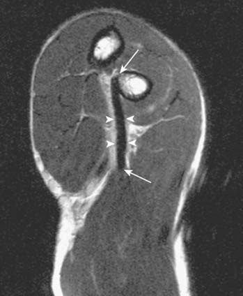
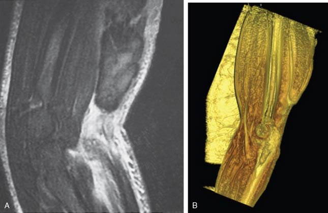
Plain x-ray study is usually normal; however, spurring of the bicipital tuberosity is sometimes seen as being suggestive of a chronic tendon enesiopathy. Magnetic resonance imaging is commonly used to make or to confirm the diagnosis,47 especially when the lacertus fibrosus is intact and the typical retraction deformity is not present or when trying to differentiate complete tears from partial tears. The treatment of complete tears without retraction or partial tears can benefit from precise delineation of the extent of the pathology. Guiffre and Moss26 have described the Flexion Abduction Supination (FABS) view. With the elbow flexed and forearm supinated, the radial tuberosity is directed medially and the distal biceps tendon is almost a direct line from the tuberosity to the muscle belly (Fig. 34-8), allowing a longitudinal view to include the muscle belly, tendon, and the difficult-to-assess bicipital tuberosity insertion (Fig. 34-9). Because the tendon is assessed longitudinally and is at full length and partially unravelled, the differentiation of partial from complete tears is made easier (Fig. 34-10). In addition, the elbow is near the center of the magnet and, thus, fat suppression is optimal, enhancing visualization of small amounts of fluid. Continuing technical advances allow visual reconstruction and enhancements (see Fig. 34-10B).
Surgical Findings
If the biceps tendon is explored early, local hemorrhage is present in the antecubital space but usually is not extensive. The tendon may have recoiled into the muscle or may lie loosely curled in the antecubital fossa. Invariably, the separation is clean and from the radial tuberosity,8,21,46,52,57,61 with a bulbous stump of degenerative tendon. This supports our histologic studies and the hypothesis that the underlying lesion is degeneration at the site of attachment.14,18 After several months, typically the tendon has retracted into the substance of the biceps muscle and cannot be retrieved to be reattached. However, this response as well as the status of the lacertus fibrosus is variable.
TREATMENT
Acute Disruption
Reliance on the older literature is unwise. Nonoperative management has been reported to provide satisfactoryresults37; however, the functional superiority of surgical treatment is obvious when the results of cases treated with and without surgical intervention are reviewed.57 The recent literature offers overwhelming documentation of the excellent results of early repair.2,6,50,60 Although tendon strains will heal, true partial rupture does not and chronic pain necessitates surgical repair.
Surgical Repair
Reattachment to the radius by any one of several techniques5,9,24,59,69 is clearly the treatment of choice. The difficulty of performing the anterior exposure needed to avoid radial nerve injury has inspired the development of a second incision that is placed over the dorsal aspect of the forearm.12 Curiously, this technique appears first to have been employed in 1937 by Plummer to reattach the biceps tendon to the proximal ulna.21 It is paramount to understand that the two-incision Boyd-Anderson technique originally described has been modified at the Mayo Clinic to reduce the likelihood of the development of ectopic bone between the radius and ulna. This point is explained later.
Because of concern over the development of ectopic bone associated with the two-incision technique, and with the advent of suture anchors, the anterior exposure using these anchors is gaining popularity. If the procedure is performed promptly, the tract of the biceps tendon is still present and is easily identified. Later (more than 2 weeks after injury), the tract may be obliterated, making the exposure more difficult36 and complications more frequent.44
SURGICAL TECHNIQUE
Two-Incision Technique (Mayo)
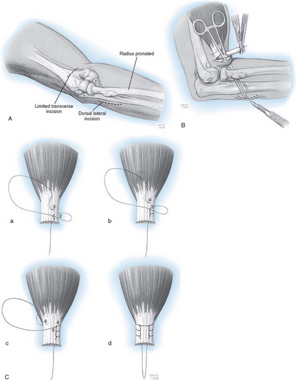
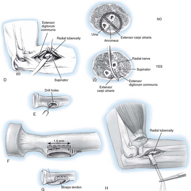
This technique has been described in detail elsewhere.56 With the patient in the supine position, a tourniquet is applied to the arm and the extremity is prepared, draped, and placed on an elbow table. A limited 3-cm transverse incision is made in the cubital crease (Fig. 34-11). The brachium is grasped and milked distally to deliver the biceps tendon. In the majority of cases, the tendon is readily retrieved with this maneuver. The tendon is inspected and invariably is found to be cleanly avulsed from the radial tuberosity. The distal 5 to 7 mm of degenerative tendon is resected, and two No. 5 nonabsorbable Bunnell or whipstitch (Krackow) sutures are placed in the torn tendon for a distance of at least 3 cm (see Fig. 34-11). The tuberosity is palpated with the indexfingers, and a blunt, curved hemostat is carefully inserted into the space previously occupied by the biceps tendon. The instrument slips past the tuberosity and is advanced below so its tip may be palpated on the dorsal aspect of the proximal forearm (see Fig. 34-11). A second incision is made over the instrument. The tuberosity is exposed by a muscle-splitting incision, with the forearm maximally pronated.
Note: The ulna is never exposed.24 A high-speed burr is used to evacuate a defect 1.5 cm wide and 1 cm deep in the radial tuberosity (see Fig. 34-11). Three holes are then placed 7 to 8 mm apart and at least 5 mm from the edge of the excavation. The tendon is delivered through the second incision, and No. 5 sutures are brought through the holes in the tuberosity. The tendon is carefully introduced into the excavation formed in the tuberosity, and with the forearm in the neutral position, the sutures are pulled tight and secured. The wounds are closed in layers, and a suction drain is inserted in the depths of the wound, both anteriorly and posteriorly. The elbow is placed in 90 degrees of flexion with the forearm placed in 45 degrees of supination. A compressive dressing is applied.
Stay updated, free articles. Join our Telegram channel

Full access? Get Clinical Tree


