, İsmail Demirkale1 and Cemil Yıldız1
(1)
Orthopedics and Traumatology, Mevki Military Hospital, Ankara, Turkey
Abstract
Developmental dysplasia of the hip (DDH) is one of the most common disorders of the hip and refers to a disease involving both the acetabulum and proximal femur. Early and accurate diagnosis is of paramount importance to avoid late sequel of the hip joint. To date, hip ultrasound (US) is the preferred diagnostic tool. The favored US technique is the Graf method. Features of the bony roof, osseous rim, and cartilaginous roof together with angles and age of the infant all designate the type of the hip joint. DDH has five main types with additional subtypes, and first two of those (types I and II) are described as centered hips, whereas others (types D, III, and IV) are decentered hips. For centered joints with inadequate maturation including type IIa(−), type IIb, and type IIc stable hips, a device should be used to place the hips in crouching position until appropriate maturation. Decentered joints should be reduced by a harness in the first 3 weeks of harness application. Reduction can be monitored by anterior US scanning of the hip.
Learning Outcomes
The most important reference point of a sonogram is lower limb of os ilium.
Just a standard plane of US can obtain a reproducible ultrasonographic view.
At the standard plane, the silhouette of iliac bone should be parallel to the probe.
Three landmarks should be well visualized on a sonogram: lower limb of the ilium at the depth of acetabular fossa, the midpoint of acetabular roof, and the labrum.
The examiner has to check the classical shape of chondro-osseous junction to be in proper silhouette to rule out any tilting effect.
The alpha angle quantifies the bony socket, whereas the beta angle quantifies the cartilaginous acetabular roof.
There are five types: types I and II describe the centered joints, and type D, type III, and type IV describe the decentered joints.
A newborn hip that will become a type 1 mature hip should have at least 50° of alpha angle at birth.
Premature infant should be classified according to his or her calendar age.
Type I depicts a mature hip with a well-developed socket (α ≥ 60°) and an angular bony rim.
Type IIa is subject to physiological immaturity.
Type IIb means that bony socket is deficient (α = 50–59°), and the baby is older than 12 weeks of age.
Type IIc is subject to “decentering phenomenon”; bony rim has lost its rotundity, and bony roof is severely deficient (α = 43–49°).
The main difference between a type IIc and type D hip is the β angle (type D, β > 77°).
The position of the perichondrium leads to a distinction between type III and type IV; type IV has a perichondrium toward to the acetabulum.
Two hard copies, one of them is free of figured lines, and report of the sonogram including age, hip type, alpha and beta angles, and management strategy suggestion should be included for documentation.
The recommended management of type IIa(−) hips is an abduction orthosis especially in newborn girls.
Hip reduction can be assessed by an anterior approach such as the Suzuki method by measuring the femoral head coverage and reduction quality without weaning off the harness.
Clinical Relevance
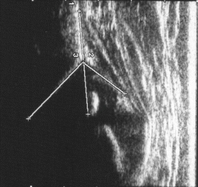
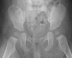
A 5-week-old female infant was brought to outpatient clinic for suspicion of left-sided developmental dysplasia of the hip according to pediatricians’ initial physical examination. On physical examination, the infant had a hip abduction asymmetry with a positive Barlow and Ortolani tests at the involved side. Immediately, lateral hip scanning was performed on the infant through the Graf method on a cradle while she was in lateral decubitus position. The hip was defined as centered hip; the bony rim was flattened (Fig. 9.1). The alpha and beta angles were measured, and it was found that bony roof was severely deficient (α = 49°) and beta angle was 59°. The hip was ultimately classified as type IIc according to the Graf classification. Thereupon, a Pavlik harness was applied, putting the involved joint in 100° of flexion by tightening the anterior straps of the harness and in moderate abduction. At 1-week intervals, follow-up was scheduled, and at the end of 3 and 6 weeks, hip ultrasound was repeated. Pavlik harness was discontinued when the acetabulum demonstrated a mature hip (α = 61°), and then the baby was put in an abduction brace for additional 6 weeks. The x-ray examination after 8 months of follow-up revealed a 25° of acetabular index angle (Fig. 9.2).


Fig. 9.1
Initial US image of the baby

Fig. 9.2
Pelvis x-ray of the same child showing sufficiently developed acetabulum
Recently, a well-done hip ultrasound is the gold standard in diagnosing hip pathologies in infancy [1]. An orthopedic surgeon should know at least main principles of lateral and anterior ultrasound scanning of an infantile hip and also can make a conclusion on an ultrasound image. The silhouette of iliac bone should be parallel to the probe, lower limb of the ilium at the depth of acetabular fossa, and the midpoint of acetabular roof and labrum should be clearly seen [2]. There are five types: types I and II describe the centered joints, and type D, type 3, and type IV describe the decentered joints.
All decentered hips including type D, type III, and type IV together with a centered but type IIc unstable hip should be reduced. Almost all type IIa(+) hips will spontaneously normalize until the end of 12 weeks of age [3]. However, the incidence of type IIa(−) hips through pursuance of type IIa hips is reported to be 15.6 %; thus, the recommended management of such hips is an abduction orthosis rather than wait-and-see regimen especially in newborn girls than boys [4].
The preferred treatment method in this age is application of Pavlik harness [5]. Follow-up through lateral scanning by the Graf method enables monitoring of the ossification of the acetabulum in centered hips. Many of Graf type III and type IV hips will reduce, and reduction quality can be checked through measurement of ischial limb-femoral distance by anterior scanning [6].
Introduction
Developmental dysplasia of the hip (DDH) is associated with specific suboptimal development affecting both the acetabulum and proximal femur, suggesting the need for concentric reduction for an acceptable hip development. Early diagnosis is the mainstay of treatment, and the gold standard for the diagnosis of DDH before 4 months of age is hip ultrasonography (HUS) especially in babies with positive risk factors [7].
Reinhard Graf was the one who first time introduced the use of HUS in screening and monitoring DDH in the late 1970s, and thereafter, HUS was widely accepted as a diagnostic tool in many countries [2, 8]. Following that, the technique has shown a progressive evolution, and Harcke, Terjesen, and Suzuki all described their own techniques [9–12]. All these HUS techniques intend to make an accurate and early diagnosis of DDH and to guide the treatment with having difference regarding image plane, direction of visualization, or focusing on morphology or stability. In the Graf method, the baby is placed on a cradle in lateral decubitus position, and the hip is scanned laterally, whereas in the Suzuki method, the baby is placed in supine position, and an anterior approach is used. The Harcke method uses a lateral approach on a baby in supine position and by applying the Barlow stress test; the hip is assessed both in neutral position and in flexion. Eventually, it’s a dynamic four-step method and involves dynamic examination of the hip both in lateral and coronal views without measurements.
Morin et al. modified the Harcke method and measured the percentage coverage of the femoral head on a lateral coronal view in slight hip flexion [12]. The Terjesen method is also a modification of the Morin method in which head coverage is measured with the aid of a baseline parallel to the long axis of the laterally placed transducer, and it is different from the Morin method with regard to figured baseline which is parallel to the lateral iliac line. The Suzuki method intends to visualize both hips via an anterior approach by a caudally directed scanning plane to transect the center of the hip. The standard plane of this method is obtained when both pubic bones are displayed on the same view with the infants’ hips in extension and then two lines are drawn. A (P) line is drawn along the anterior surface of the pubic bones, and a second line (E) is drawn from the lateral margin of the pubic bones. With a dislocated hip, femoral head crosses the P line, and in such case, the examination is repeated in abduction and flexion position. The transverse-neutral and coronal-flexion views can be combined to both detect direction of the displacement and evaluate acetabular dysplasia [13].
These four well-known procedures have their own advantages and disadvantages, but one must have in mind that a duly performed technique is the best whether an anterior or lateral technique was chosen. Among these, the Graf method is the most widely used for accurate and early diagnosis of a DDH with a well-defined standardization, and recent reports suggest the use of an anterior approach in management and follow-up of the patients with DDH [6].
Usually, suspicion of DDH begins with the very first physical examination of a pediatrician, and they consult babies with risk factors or having positive examination findings to pediatric orthopedic surgeons. According to one report, clinical examination alone in detecting DDH has a low specificity, so to prevent a misjudgment on the management of DDH, certification on the hard copy of a hip ultrasound image has a paramount importance [14]. That being said, in many cases, errors made in diagnosis or late diagnosis can lead to severe catastrophic results with a miserable lifetime quality of the patients [15]. Therefore, efforts to identify a baby with DDH, appropriate treatment, and prevention of hip dysplasia must, of necessity, become a major precedency of orthopedic surgeons.
To date, a well-done HUS is the gold standard in diagnosing hip pathologies in infancy [1]. The magnetic resonance imaging and computerized tomography can be used, but they are performed under anesthesia during infant age. Also radiation exposure is excessive at BT. Arthrography is useful but it is an invasive method. HUS visualizes chondral parts of the hip joint excellently. Currently, both radiologists and orthopedic surgeons perform HUS in these infants to screen DDH. Although a limited number of orthopedic surgeons perform HUS, more of them deal with the treatment of DDH. Thus, whether HUS is performed or not, an orthopedic surgeon should be very competent on the anatomy of newborn hip, know at least main principles of lateral and anterior ultrasound scanning of an infantile hip, and also can make a conclusion on an ultrasound image.
The Graf method has quantitative classification system and measures two angles, α and β; the Harcke and Terjesen methods determine the amount of the femoral head coverage by the osseous part of the acetabulum.
Ultrasonographic Anatomy of Newborn Hip Joint
The perichondrium of the cartilaginous acetabular roof, joint capsule, and acetabular labrum are echolucent structures, which means that the ultrasound beam can transmit through these structures. The proximal perichondrium is composed of the perichondrium, insertion of the capsule, and reflex head of the rectus femoris.
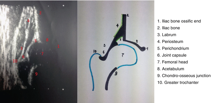
However, the bony parts also exist in the newborn hip. These are chondro-osseous junction between the shaft and neck of the femur, lower limb of os ilium and os ischium, and upper part of os ilium just above the acetabulum, and these structures are seen as bright echoes leaving an acoustic shadow behind them. The most important reference point of a sonogram is the lower limb of os ilium and the hypoechoic triradiate cartilage that exists caudal to the lower limb (Fig. 9.3) [2]. When the examiner detects the lower limb of the ilium, then he/she has to move proximally to find out the point where the concavity of bony acetabulum turns to convexity of the ilium. This point is called as “osseous bony rim,” which is the most lateral point of bony socket, and later it will be used to measure the cartilage roof angle.


Fig. 9.3
Anatomical structures of infant hip: (1) Iliac bone ossific end, (2) Iliac bone, (3) Labrum, (4) Periosteum, (5) Perichondrium, (6) Joint capsule, (7) Femoral head, (8) Acetabulum, (9) Chondro-osseous junction and (10) Greater trochanter
< div class='tao-gold-member'>
Only gold members can continue reading. Log In or Register to continue
Stay updated, free articles. Join our Telegram channel

Full access? Get Clinical Tree


