Fig. 23.1
Arthroscopic view of meniscus remnant impinging between the femoral implant and the inlay
With the development of mobile bearing inlays, fat pad impingements were reported, for example, in a case series of 218 patients (230 knees) with a PCL-retaining TKR (A/P Glide LCS DePuy AG, Cham, Switzerland) [10]. Typical symptoms such as anterior knee pain and tenderness of the anterior soft tissue to palpation were reported in 18.7 % of the patients. Revision surgery was successful and included fat pad and PCL resection as well as exchange of the tibial inlay for a rotating-platform bearing inlay.
Fat pad impingements were reported in PCL-retaining TKR (A/P glide). Typical symptoms were anterior knee pain and tenderness to palpation.
23.1.2 Patellofemoral Soft-Tissue Impingement
Patellofemoral impingement has been extensively described in Chap. 42.
23.2 Bony Impingement
Bony impingement includes several different impingement types such as posterior femoral bony impingement, bony patellofemoral impingement, patellotibial impingement, and bony anterior impingement.
Bony impingement can be differentiated into posterior femoral, patellofemoral, patellotibial, and anterior bony impingement.
23.2.1 Posterior Femoral Bony Impingement
Posterior femoral bony impingement can be caused by residual osteophytes or substantially exposed bone proximal to the posterior shield of the femoral implant. Here, impingement occurs with the tibial inlay during deep knee flexion [11, 12]. The size of the posterior osteophytes correlates with the decrease of maximum knee flexion [13]. Using a geometric model, osteophytes with a radius of below 2.9 mm allowed 120° flexion, and osteophytes with more than 6.5 mm allowed 105° before impinging. These results were in vivo confirmed in a study evaluating 92 patients using a low-contact rotating platform TKR. It showed the negative effect of osteophytes on impingement and range of motion [12]. Unfortunately, even properly removed osteophytes during the primary TKR can regrow and then may damage the tibial inlay as shown in a case report 8 years after TKR [14]. A further risk factor for posterior femoral bony impingement, and far more common, is a too small femoral TKR implant [13].
Posterior femoral bony impingement can be caused by residual osteophytes, substantially exposed bone proximal to the posterior shield of the femoral implant and small femoral implants. The size of the posterior osteophytes determines the decrease of maximum knee flexion.
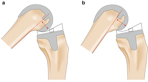
The size of the femoral implant of the TKR also plays an important role for the posterior condylar offset (PCO) [15] (Fig. 23.2). It was postulated that a decreased PCO leads to earlier impingement and less range of motion (Fig. 23.2).


Fig. 23.2
This schematic drawing illustrates the effect of the size of PCO and femoral implant size. (a) Shows a limited knee flexion angle due to a small PCO in a small femoral implant. (b) Shows a bigger PCO and a bigger femoral implant resulting in an increased flexion angle
A decrease in posterior condylar offset (PCO) can lead to decreased flexion. This only occurs in PCL retaining TKR. The amount of PCO depends strongly on the size of the femoral implant with respect to the size of the distal femur.
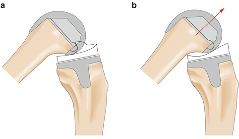
In a finite element model without soft tissue, downsizing the femoral implant from size five to four in PCL-retaining TKR decreased maximum knee flexion from 135 to 120°, because of less PCO of the smaller femoral implant [13]. Interestingly, the effect of PCO and impingement seems to depend on the type of TKR. For posterior-stabilized (PS)-TKR designs, no correlation was found between PCO and range of motion and thus impingement [11]. In contrast using PCL-retaining TKR, there is a correlation between PCO and range of motion as well as impingement [15]. In an in vivo study with 179 patients, a decreased PCO lead to limited flexion and earlier impingement. One millimeter less PCO resulted in a reduction of 6° of flexion. Thus, downsizing of the femoral implant should be avoided, because it endangers bone overlapping and thus increased PCO with impingement and less range of motion. Unfortunately, downsizing happens easily because many systems reference the implant size at the anterior femur. The reason for the vulnerability of PCL-retaining TKR might be the roll-forward mechanism of the femoral implant during knee flexion, which leads to an earlier onset of impingement and less range of motion (Fig. 23.3) [15–20]. This fact was confirmed by a study implanting bilateral TKR, one knee with a PCL-retaining and one knee with a PS-TKR [21, 22]. Knees with a PS-TKR showed significantly greater knee flexion angles compared to knees with a PCL-retaining TKR. Patients with a decreased PCO in the PCL-retaining group had a lower range of motion. In contrast, in the PS-TKR group, the magnitude of the PCO did not influence the range of motion. Interestingly, newly advocated high-flexion TKR designs have not yet shown significant improvement in range of motion compared to standard PS-TKR (106 ± 14° vs. 105 ± 13°, respectively) [23].


Fig. 23.3
This schematic drawing illustrates the effect of an anterior femoral translation. The more the femur translates anteriorly during knee flexion, the higher is the chance of impingement between the femur and the tibial inlay. (a) No anterior translation, (b) anterior translation and thus limited knee flexion
23.2.2 Bony Patellofemoral Impingement
Another type of bony impingement is the bony patellofemoral impingement between the patella and the femoral implant of the TKR, which shows similar symptoms when compared to the soft tissue patellofemoral impingement. Patients complain about locking sensations, especially during knee extension. Important for diagnostics besides history and physical evaluation are radiographs. In additional to standard radiographs such as lateral and anterior-posterior knee views, a specific patella radiograph should be taken, preferably a weight-bearing view such as the sunrise view or Merchant’s view, the modified Merchant’s view [24], the patella axial view (45°), or patella defile views [25, 26]. Weight-bearing views are especially important to detect impingement of the medial patellar facet, which correlates highly with the severity of pain [25]. In contrast, lateral patellar impingement does not seem to correlate with the severity of pain, neither in weight-bearing nor in the non-weight-bearing Merchant’s views [25]. Lateral knee radiographs are important to assess the patella position in relation to the joint line. Patella baja is one most common causes creating patellofemoral impingement [27, 28]. Impingement between the lateral facet of the patella and the femoral implant may also occur when the patellar implant is placed too far medially or the tibial implant internally rotated [26]. A case series of 19 TKR patients suffering anterior knee pain all showed lateral patellofemoral impingement. A combined lateral and medial patellofemoral impingement was seen in three of them [29]. This cohort represented 7 % of all painful TKR revised. Patients were treated operatively with facetectomy or revision of the patellar implant. At 1-year follow-up, all patients had significant improvement in the Knee Society Score. Some authors discussed a patellar chamfering in case of a mismatch between the patellar prostheses and the retropatellar area [30].
The bony patellofemoral impingement occurs between the patella and the femoral implant of the TKR. Typically patients complain about locking sensations, especially during knee extension. Patella baja is one of the most common causes.
23.2.3 Patellotibial Impingement
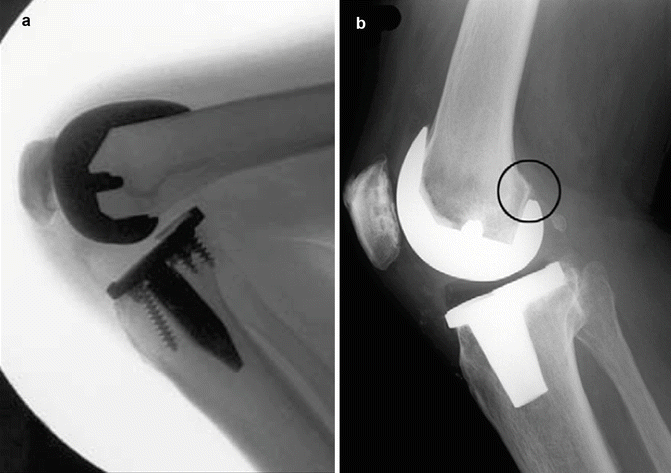
The patellotibial impingement is another type of bony impingement. The patella impinges against the tibial implant or the tibial inlay. This impingement occurs during knee flexion caused by joint line elevation or patella baja [31–34]. Patella baja, due to joint line elevation (four out of seven knees in the abovementioned case series), should rather be called “pseudo-patella baja” (Fig. 23.4). A pseudo-patella baja may occur due to over-resection at the distal femur or due to the usage of thicker polyethylene inlays in order to achieve mediolateral stability. Low (distal) positioning of the patellar implant may also cause impingement.


Fig. 23.4
These lateral radiographs show two typical reasons for impingement: (a) a missing anterior translation of the femoral implant, and (b) an osteophyte proximal at the dorsal aspect of the femoral implant
Furthermore, patella baja can develop postoperatively as a result of local or general arthrofibrosis [34, 35]. The main symptoms of patellotibial impingement are pain and reduction in range of motion. Lateral radiographs in maximum flexion will help to diagnose impingement because, after several months, signs of erosion of the inferior patella pole might be visible.
In patellotibial impingement, the patella impinges against the tibial implant or the tibial inlay. It occurs in deep knee flexion, mainly in patients with a patella baja.
23.2.4 Fabella Impingement
Fabella impingement and impingement after posterolateral cement extrusion are very rare types of impingement. Fabella impingement occurs between a large posterolateral fabella and the posterior rim of the tibial inlay [36] or the femoral implant [36–38]. Patients feel pain and in some cases a painful click during knee flexion at about 90° with exacerbation during plantar flexion of the foot [37]. Treatment of choice is a removal of the fabella through a posterolateral approach. Posterolaterally extruded cement from the tibial implant can impinge against the fibular head and cause pain and ROM deficit [39].
Fabella impingement and impingement after posterolateral cement extrusion are rare.
23.3 Implant-Related Impingement
Post-cam impingement occurs between the posterior aspect of the tibial inlay and the cam of the femoral implant. It is particularly found in deep knee flexion and can result in severe wear or even fracture of the tibial post [40–42]. Post-cam impingement occurs in PS-TKR design due to the rollback mechanism of the femur during deep flexion. High-contact stresses (more than 30 MPa) were measured at the posterior aspect of the tibial post. These high-contact stresses exist independently of the PS-TKR designs [40] and thus, in almost 100 % of the knees, wear of the post happens [43–46]. However, the design of the post influences the amount as well as the characteristics of the wear [46–49]. In general, two different post designs can be distinguished: (1) flat on flat and (2) curve on curve. The flat-on-flat design has flat contact surfaces at the tibial post and at the cam; the curve-on-curve design has curved contact surfaces at the tibial post and at the cam [47]. The latter one was found to decrease the stress concentration at the tibial post during knee flexion. It also decreases edge loading on the tibial post [47, 48]. The curve-on-curve design is beneficial in knee kinematics in terms of internal rotation in deep knee flexion [49]. Some studies mentioned that impingement could be avoided by an implantation technique such as excessive posterior tibial slope [44, 50, 51].
Post-cam impingement occurs between the posterior aspect of tibial inlay and the cam of the femoral implant. Typically it occurs in deep knee flexion. The design of the post and cam influences the stress on the implant.
23.3.1 Anterior Implant Impingement
Anterior implant impingement occurs between the anterior aspect of the tibial post and the femoral implant box and can result not only in wear at the anterior but also at the medial and lateral sides of the tibial post [45]. It typically occurs during hyperextension and in low flexion angles [3]. The incidence of anterior wear was found to be between 36 and 88 % [3, 52, 53]. Severe wear was found in up to 30 % of the cases [45]. The anterior tibial post functions as an ACL during hyperextension in PS-TKR [54]. Further risk factors for anterior post impingement are increased dorsiflexion of the femoral implant and increased posterior tibial slope. Anterior implant impingement is not specifically linked to a certain TKR design; however, the PS-TKR design seems, in particular, to be prone to anterior implant impingement [51]. In a dynamic in vivo three-dimensional fluoroscopic gait analysis of PS-TKR knees, anterior implant impingement was observed in all patients during stance phase in hyperextension (on average 12°) [51]. A static in vivo study found an anterior impingement in 63 % of the patients with PS-TKR implants at full extension. However, this study evaluated impingement during a single stand in a quasi static condition [55]. Anterior impingement was also associated with higher degrees of knee extension and increased femoral implant flexion.
Stay updated, free articles. Join our Telegram channel

Full access? Get Clinical Tree


