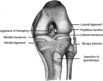
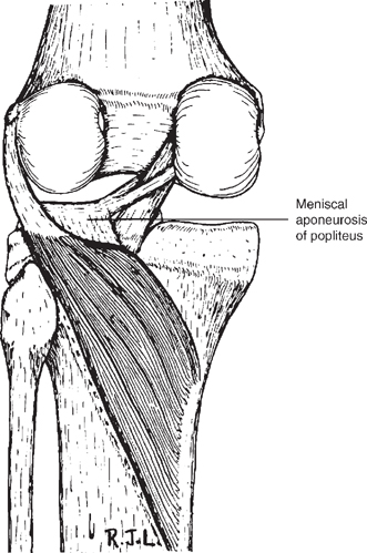
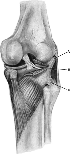
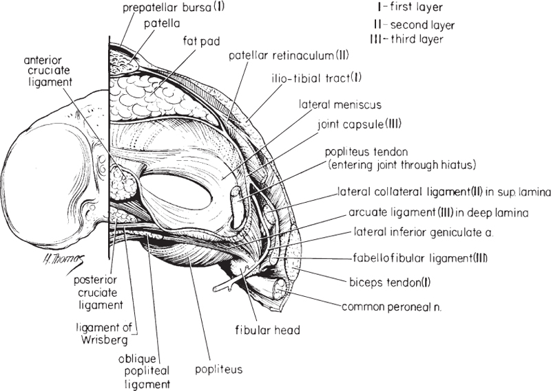
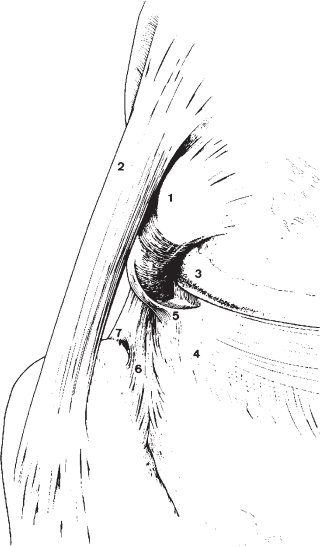
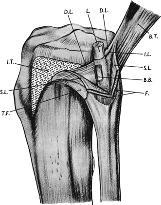
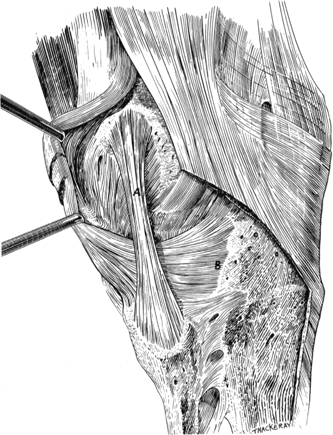
2 It is well recognized that the anatomy of the posterolateral corner of the knee is more complex than that on the medial side. This is partly due to the developmental phylogeny of the knee that has evolved between species over time. In lower animal species, the fibula articulates with the femur, and it has descended over eons to articulate with the tibia in higher order animal species.1–5 Previous studies have reported that in lower animal species, there is a meniscus between the articulation of the femur and the fibular head.1,2,4–6 Many authors have theorized that this femorofibular meniscus evolved into the popliteus tendon of the knee.1–5 In addition to these changes that have been noted in the popliteus complex, the biceps femoris complex in other animal species has attachments around the knee that are significantly different from those found in humans.4,6–8 It has also been reported that the human knee is the only one that has an iliotibial band over the lateral aspect of the knee.7,9 The bony anatomy of the lateral aspect of the knee is also an indirect contributing factor to the complexity of its anatomy. Although the medial femoral condyle, with its convex surface, and the medial tibial plateau, with its concave surface, have two opposing surfaces that are inherently stable as they cup together, one can see that this is not true for the lateral compartment of the knee. Both the lateral femoral condyle and the lateral tibial plateau have convex surfaces that make the bony geometry inherently unstable.10,11 The two convex surfaces rely on point contact to provide bony stability to the lateral compartment of the knee. This inherently unstable bony geometry places increased reliance on the integrity of the multiple individual soft tissue structures of the posterolateral aspect of the knee to provide static stability for the lateral side of the knee (Fig. 2-1). In addition, when the posterolateral structures of the knee are injured, there is less ability for these structures to remain in opposition, so there is less ability for these structures to heal. The difference in the inherent stability of the medial and lateral menisci also contributes to differences in the inherent stability and healing potential between the two tibiofemoral compartments. The medial meniscus has a more stable meniscotibial attachment and has less relative motion across the medial tibial plateau with flexion and extension of the knee. However, the lateral meniscus is much less stable and has a less restraining meniscotibial attachment, which results in far greater motion of the lateral meniscus with knee motion than its medial counterpart.12 The increased lateral meniscal motion further accentuates the unstable bony geometry of the lateral compartment of the knee. In addition to the confusion about posterolateral knee anatomy from its developmental phylogeny and bony anatomy differences, there has also been a great deal of confusion created by varying anatomic descriptions of these structures by anatomists and physicians studying these structures. Over the last century and a half, several published articles have described the anatomy of the posterolateral knee using varying nomenclature, descriptions, and illustrations. The variations in nomenclature used in different reporting institutions have especially contributed to the confusion regarding the anatomy of the posterolateral corner of the knee. A careful review of the literature reveals that a lot of the confusion about the nomenclature of posterolateral knee structures may be due to the revision of the standard anatomic nomenclature by the International Congress of Anatomists late in the first half of the 20th century. Their combined efforts to update the nomenclature of the knee resulted in the Nomina Anatomica,13 which attempted to modernize previous anatomic descriptions. As one author from that time pointed out,14 however, the short external lateral ligament (which appears to be the popliteofibular ligament in his descriptions) was omitted from the nomenclature revision. As a result of this omission, this structure has not been specifically depicted or described well in anatomic textbooks since that point in time. It appears that a lot of the confusion about the nomenclature of the posterolateral knee is due to this omission, because varying authors have attempted to describe this structure in many different ways over the past 50 years. In fact, other authors who have quoted Last14 since his publication in 1950 seem to be describing the short external lateral ligament as a different structure than what he had originally described it to be.4,15,16 A review of these previous studies and a scholarly interpretation of which structures they may be referring to are presented on a structure-by-structure basis in this chapter in an attempt to resolve this confusion and to present the case for a descriptive anatomic nomenclature basis for the posterolateral structures presented in Chapter 3. It is important to understand that a unified nomenclature for the posterolateral knee is essential to elucidate the contributions of each structure to knee stability and joint function, and to accurately compare anatomic, biomechanical, clinical, and radiographic imaging studies. In one of the early studies in the English-language literature on the anatomy of the posterolateral knee, Sutton17 reported in 1884 on the anatomy of the “external lateral ligament of the knee joint,” which we now know to be the fibular collateral ligament. Sutton described it as extending over the lateral aspect of the knee, and he theorized that this ligament was an extension of the peroneus longus muscle and tendon to the knee. He felt that it was an extension of these structures due to his comparative anatomy studies in other animal species. These species included the opossum, domestic ox, and the gibbon. Figure 2-1 Lateral ligament (fibular collateral ligament) of the knee joint coursing from the lateral femoral condyle to the fibula with the biceps femoris enveloping the fibular attachment. (From Last RJ. Some anatomical details of the knee joint. J Bone Joint Surg 1948;30B:683–688, with permission.) Last18 reported in 1948 that the lateral ligament of the knee joint of “current” British terminology was represented by the long external lateral ligament of “old terminology” (Fig. 2-1). He reported that it originated from the lateral epicondyle of the femur and inserted on the fibular head. Last also observed that the tendon of the biceps femoris wrapped around the posterior aspect of the lateral ligament of the knee. In an article on the prenatal development of the human knee, Gray and Gardner19 reported that the fibular collateral ligament was present in a 9-week-old fetus as a thin cellular band that descended from the lateral aspect of the femoral condyle to the fibular head. By 14 weeks of age in the fetus, the fibular collateral ligament resembled that of the adult knee. They reported that their findings in the fetus indicated that the fibular collateral ligament developed in situ, and that there was no evidence to support Sutton’s17 theory that the fibular collateral ligament was an extension of the peroneus longus fascia over the lateral compartment of the leg. Kaplan20 in 1957 reported that the most important structure found under the fascia on the lateral side of the knee was the lateral collateral ligament. He reported that this ligament coursed between the lateral condylar tubercle, which we now call the lateral epicondyle, of the femur and the middle of the lateral surface of the fibular head. To understand the complex anatomy of the popliteus complex, it is very important to review both older human literature and comparative anatomy studies. It is especially clear that this portion of the anatomy of the posterolateral knee has been one of the most confusing for clinicians to understand. A large part of this confusion is due to the fact that the popliteus attachment to the fibular head (the popliteofibular ligament) was omitted from much, but not all, of the English-language orthopaedic literature for much of the latter part of the 20th century. Those articles that recognized this structure were in more obscure orthopaedic or anatomy journals that went unread by most practicing orthopaedists. Therefore, many different descriptions with varying nomenclature of the structures in this area emerged over time. Higgins21 reported that the proximal attachments of the popliteus complex were to the lateral femoral condyle and the fibular head. The fibular attachment was noted to consist of two strong and distinct bands. In addition, Higgins noted attachment fibers from the popliteus to the posterior joint capsule, the coronary ligament to the lateral meniscus, and the lateral meniscus itself. The femoral attachment of the popliteus was noted to be in a groove on the femur and distal and anterior to the “long external lateral ligament” (fibular collateral ligament). The popliteus tendon was also noted to form a groove where it crossed the posterolateral aspect of the lateral tibial plateau. We now know that his studies described, in an accurate qualitative manner, the anterior and posterior divisions of the popliteofibular ligament, the popliteomeniscal fascicles, and the popliteal aponeurotic attachments to the lateral meniscus. Fürst1 reported in the German literature in 1903 that the tendon of the popliteus and its muscle were two structures that developed independently in the animal world. He reported that the popliteus muscle attached directly to the fibular head in lower animal species. These lower animal species were also noted to have a meniscus between the fibula and femur. The popliteus tendon was felt to have evolved from this femorofibular meniscus. In humans, Fürst reported that the popliteus muscle had two proximal attachments, with one on the lateral femoral condyle (the popliteus tendon attachment to the femur) and the other on the fibular head (the popliteofibular ligament). Fürst also noted that the popliteus tendon created a small sinusoidal cartilage indentation (which Stäubli and Birrer22 noted as the sulcus statorious of Fürst) on the border of the lateral femoral condyle of the knee near full extension. Fürst felt that the sulcus statorious was acquired due to the upright posture of humans and that the popliteal sulcus on the lateral femoral condyle evolved due to the flexed knee position in gait of most lower animal species. Taylor and Bonney2 reported that in reptiles the popliteus muscle originated from the fibular head and also had a small attachment to the femorofibular meniscus. This femorofibular meniscus attached to the lateral femoral condyle via a femorofibular ligament. In addition, they noted that some lower animals and marsupials had a popliteus muscle that attached to the fibular head. However, in higher marsupial species such as kangaroos, and in most higher mammals, the popliteus acquired its femoral attachment by the development of the femorofibular meniscus, and its femorofibular ligamentum attachment, to evolve to form the popliteus tendon femoral attachment. The authors also noted that in higher mammals, in which motion between the tibia and fibula were more important, the popliteus attained a firm attachment to the fibular head. We now know that this structure is the popliteofibular ligament. Haines6 also performed extensive comparative anatomy studies of the posterolateral aspect of the knee. He reported that in the crocodile, which appears to have the most primitive type of knee joint in currently living animals, the popliteus muscle attached directly to the fibular head. In marsupial species, the femorofibular meniscus was a pad of fibrocartilage that articulated with and was attached to the lateral femoral condyle and fibula. This meniscal structure had an attachment to the popliteus muscle and coursed in a groove on the lateral aspect of the lateral femoral condyle when the knee was in maximal flexion. He theorized that this femorofibular meniscus evolved over time to form the popliteus tendon and its attachment on the femur. Last14 reported that the popliteus complex emerged from underneath the “arcuate ligament” and that a very substantial portion of the popliteus muscle arose not from the tendon of the popliteus but from a portion of the arcuate ligament. A careful review of his illustrations demonstrates that the arcuate ligament that he was referring to was the popliteal aponeurosis to the lateral meniscus (Fig. 2-2). Last suggested that this attachment of the popliteus to the posterior aspect of the lateral meniscus helped to protect the lateral meniscus from injury. He also noted that fibrous strands directly connected the popliteus tendon to the lateral meniscus by synovial reflections above and below the meniscus. These are now known as the popliteomeniscal fascicles. In addition, he noted the popliteus tendon made a narrow groove in the posterolateral aspect of the lateral tibial plateau, just proximal to the fibular styloid. This bony landmark marks the location of the popliteus musculotendinous junction and is currently utilized to note the location of the popliteus tunnel for an anatomic posterolateral reconstruction (see Chapter 7). Kaplan20 reported that the popliteus tendon originated in front of and below the origin of the lateral collateral ligament (fibular collateral ligament) on the femur, and it was separated from the fibular collateral ligament by the joint capsule. He also stated that the popliteus tendon itself rarely adhered to the meniscus, and there was an intimate connection of the popliteus tendon to the fibular head by a ligament that connected both structures.20,23,24 Kaplan reported that this connection sometimes reached the tibiofibular joint and was found in other species, including the chimpanzee. In this regard, he was referring to the popliteofibular ligament. Kaplan also noted that the muscular body of the popliteus, in contrast to the popliteus tendon, adhered firmly to the lateral meniscus, the coronary ligament to the lateral meniscus, and the posterior capsule of the knee joint.20,23,24 He reported that this attachment occurred through the means of a thick fascia, which he termed the popliteal fascia. We now know this to be the popliteal aponeurosis to the lateral meniscus. The relative immobility of the popliteus tendon at the knee was also noted by Kaplan, which he felt made this tendon a static stabilizer of the knee in all positions. He also noted the rare occurrence of a sesamoid bone, the cyamella, in the tendon of the popliteus in humans.20 He also felt that the popliteus tendon was almost never adherent to the lateral meniscus (which we now know to be incorrect because of its popliteomeniscal fascicle attachments). 22,25 Kaplan4 also noted that the meniscal remnant between the femur and the fibula in lower animal species eventually evolved into the popliteus tendon. In fact, he noted that the femoromeniscal ligament to the femorofibular meniscus appeared to have evolved into the popliteus attachment to the femur, whereas the meniscofibular ligament evolved into the popliteofibular ligament. Figure 2-2 Posterior view of a left knee demonstrating the popliteus attachment to the posterior horn of the lateral meniscus (popliteal aponeurosis attachment to the lateral meniscus). (From Last RJ. The popliteus muscle and the lateral meniscus: with a note on the attachment of the medial meniscus. J Bone Joint Surg 1950;32B:93–99, with permission.) Lovejoy and Harden26 reported that the popliteus had three attachments at the knee. They described them as attachments to the lateral femoral condyle, the fibular styloid (which we now know as the popliteofibular ligament), and the posterior horn of the lateral meniscus (the popliteal aponeurosis to the lateral meniscus). The femoral attachment was in a fossa that was anteroinferior to the lateral (fibular) collateral ligament. This fossa was the popliteus sulcus. They found a fibular attachment from the popliteus muscle to be present in all specimens (popliteofibular ligament). They noted that the femoral attachment, or the popliteus tendon, and the fibular attachment (popliteofibular ligament) formed the arms of a Y-shaped ligament that they stated had been previously described as a separate entity (Fig. 2-3). They reported that this separate entity was incorrectly called the “arcuate” ligament. They felt that this was incorrect because this structure was not a distinct separate entity, but rather a distinct portion of the popliteus complex. However, their article was in the anatomy literature and thus not read by most orthopaedists, and so it was not well recognized at that time. Figure 2-3 Illustration of the posterior view of a right knee demonstrating the Y-shaped ligament described by Lovejoy. A, popliteus femoral origin; B, popliteus aponeurosis to lateral meniscus; C, popliteofibular ligament. (From Lovejoy JF, Harden TT. Popliteus muscle in man. Anat Rec 1971;169:727–730, with permission.) In 1975, Reis and de Carvalho27 described the attachments of the human popliteus muscle complex at the knee. The proximal attachment of the popliteus muscle was in a depression just below the lateral epicondyle and was beneath and anterior and medial to the fibular collateral ligament’s femoral attachment. They noted that the popliteus tendon had an oblique direction upward and downward, and formed approximately a 45-degree angle over the horizontal plane when it passed by the lateral tibial plateau. They concluded that the popliteus muscle had four attachments near the knee. They were the direct origin on the femur, the meniscal attachments, the attachments to the posterior capsule (popliteal aponeurosis), and the fibular head attachment (the popliteofibular ligament). They reported that the meniscal attachments (popliteomeniscal fascicles) united the popliteus tendon to the lateral meniscus and that the fibular attachment (the popliteofibular ligament) originated from the musculotendinous junction of the popliteus. In 1979, Cohn and Mains28 described the anatomy of the popliteal hiatus. They reported that the anatomy of the popliteus complex is constant at the popliteal hiatus. They observed two distinct ligamentous fascicles to the lateral meniscus: one for the superior surface and one for the inferior surface of the meniscus. These fascicles were noted to make up the floor and the roof of the popliteal hiatus, respectively. In carefully reviewing the authors’ illustrations, one can see that the superior fascicle corresponds to what we now know as the posterosuperior popliteomeniscal fascicle, and the inferior fascicle corresponds to the anteroinferior popliteomeniscal fascicle. Fabbriciani et al5 reported in the Italian literature on the anatomy of the popliteus muscle complex of the knee in 1982. These authors reported that the popliteus muscle continued into a complex aponeurosis consisting of popliteocapsular (the popliteal aponeurosis to the lateral meniscus), popliteofibular, and popliteomeniscal (the popliteomeniscal fascicles) fibers. They reported that the popliteus developed into a strong tendon that attached on the lateral femoral condyle. They stated that there were both superior and inferior popliteomeniscal fibers (popliteomeniscal fascicles). In addition, they summarized some of the developmental phylogeny literature. They reported that comparative anatomy studies demonstrated that in lower order vertebrates, the fibula and the tibia each had a separate articulation with the femur with its respective meniscus. They also reported that the fibers of the popliteus muscle attached directly to the fibular head in lower animal species. As the fibular head descended distally in higher animal species, the differentiation of the meniscus between the femur and fibula was noted to occur. In the later stages of phylogenic development, they postulated the popliteus muscle became attached to the femorofibular meniscus, which evolved to become the popliteus tendon, and the popliteus muscle developed a tendinous attachment to the fibular head, which evolved to become the popliteofibular ligament. In addition, the popliteus tendon maintained its ties to the lateral meniscus (which we now refer to as the popliteal aponeurosis to the lateral meniscus and popliteomeniscal fascicles). Seebacher et al15 reported that the inner (deep) lamina of layer III (of the three anatomic layers that they described in their article) terminated posteriorly as a “Y-shaped arcuate” ligament, which spanned the junction between the popliteus muscle and its tendon from the fibular head to the femur. They stated that the superficial and inner lamina of layer III were always separated from each other in the region of the fibula by the inferior lateral genicular vessels (Fig. 2-4). They noted that the “arcuate” ligament inserted at the apex of the fibular styloid and was firmly adherent to the musculotendinous junction of the popliteus. They reported that in all cases a reinforcing structure was noted to course directly from the fibula to the popliteus musculotendinous junction. Seebacher et al also noted that a deep, or inner, lamina formed the hiatus for the popliteus tendon (i.e., the popliteomeniscal fascicles). In understanding this work by Seebacher et al, it is important to note that a further anatomic study29 from their center noted, in retrospect, that a direct attachment of the popliteus complex to the fibular head had not been recognized by Seebacher et al. These later researchers reported that they now understood this description to be the popliteofibular ligament. It is very important to recognize the group’s retraction of the use of the term arcuate ligament, because the work of Seebacher et al15 is still widely quoted in the orthopaedic and radiology literature. Figure 2-4 Axial illustration of a right knee with emphasis on the posterolateral knee structures. In this depiction, the authors note that the fabellofibular ligament and arcuate ligament (popliteofibular ligament) are separated from each other by the inferior lateral genicular artery. (From Seebacher JR, Inglis AE, Marshall JL, Warren RF. The structure of the posterolateral aspect of the knee. J Bone Joint Surg 1982;64A:536–541, with permission.) The human embryonic development of the popliteus complex was described by Oransky and colleagues30 in 1989. These authors found that the attachments of the popliteus complex to the lateral meniscus and its fibular head were formed during the process of embryonic cavitation during the formation of the popliteal hiatus. They found that this occurred at about the 10th week of embryonic development and it resulted in a separation of the popliteus tendon from the lateral meniscus. The popliteofibular ligament was found to develop soon after this. Oransky and colleagues also verified the location of the popliteofibular ligament in adult human cadavers and reported that it was a strong and constantly found structure in all specimens. They noted that it coursed between the posteromedial aspect of the fibular head and the lateral aspect of the popliteus tendon. Another group of authors16 reported on the popliteofibular ligament and found it to be present in 98% of their knee dissections. They reported that it was a short, strong, tendinous band that coursed between the popliteus tendon and the fibular styloid. They also stated that they believed that this structure should be considered a true distinct ligament of the knee joint with importance in preventing posterolateral rotation of the knee. In the early 1990s, Stäubli and coauthors22,25 reported extensively on the anatomy of the popliteus complex and its attachments to the lateral meniscus at the popliteal hiatus (Fig. 2-5). They noted that the popliteus tendon attachment on the femur was at the anterior aspect of the popliteal sulcus and anterior and distal to the lateral epicondyle in all cases. The popliteus tendon also caused a sinusoidal indentation of the articular cartilage where it crossed the lateral femoral condyle in extension, and only lay within the confines of the popliteal sulcus when the knee was significantly flexed. The anteroinferior popliteomeniscal fascicle was always present and blended into the middle third of the lateral meniscus. This fascicle created the anterior floor of the popliteal hiatus that can be seen arthroscopically. The posterosuperior popliteomeniscal fascicle was also found to blend into the posterior horn of the lateral meniscus and the meniscofemoral portion of the posterior capsule of the joint. Stäubli and coauthors also described the popliteofibular fascicle (ligament), which consisted of anterior and posterior divisions. They reported that it arose from the proximal tibiofibular joint and fibular styloid region and extended to the musculotendinous junction of the popliteus. They reported the popliteofibular ligament had the shape of an inverted Y and consisted of an anterior division that blended proximally into the anteroinferior popliteomeniscal fascicle and a posterior division that attached to the posterior aspect of the fibular styloid and was adjacent and adherent to the proximal tibiofibular joint capsule. They also reported that the inferior lateral genicular artery passed posterior to the popliteofibular ligament and anterior to the fabellofibular ligament and its associated structures. Figure 2-5 The popliteus complex and its relationship to the lateral meniscus and fibula in a right knee. 1, popliteus tendon; 2, fibular collateral ligament; 3, sulcus statarius; 4, meniscotibial portion of mid-third lateral capsule ligament; 5, anteroinferior popliteomeniscal fascicles; 6, anterior division of popliteofibular ligament; 7, posterior division of popliteofibular ligament. (From Stäubli HU, Birrer S. The popliteus tendon and its fascicles at the popliteus hiatus: gross anatomy and functional arthroscopic evaluation with and without anterior cruciate ligament deficiency. Arthroscopy 1990;6:209–220, with permission.) Watanabe et al31 reported that the popliteofibular ligament was present in 93% of their dissected knee specimens. They reported that the popliteus tendon attachment on the femur was consistently present at the popliteal sulcus. They also did three-dimensional geometry studies of the popliteus tendon with varying degrees of knee flexion by inserting metal markers into the anterior and posterior aspect of the popliteus tendon and obtaining radiographs. They found that the anterior part of the popliteus tendon became tighter with higher knee flexion angles and the posterior part of the popliteus tendon became tighter near extension. From this information, they theorized that the posterior aspect of the popliteus tendon and the popliteofibular ligament would act as restraints near full extension, whereas the anterior portion of the popliteus tendon would act as a posterolateral rotatory restraint with increasing knee flexion. Maynard and coauthors29 also reported on the anatomy of the popliteofibular ligament. Their dissection technique helped to isolate it better than most previous techniques in the past. They dissected their specimens from inside the joint to outside rather than the usual outside-in technique. By this method, they were able to isolate the popliteofibular ligament better than did previous dissection techniques. These authors found the popliteofibular ligament to be substantial in size and present in all of their dissected knees. The popliteofibular ligament fibers were noted to fuse to the popliteus tendon and to diverge to the proximal and posterior aspect of the fibular head. These authors were noted to be from the same research center as Seebacher et al15 and Gollehon et al,32 and they were able to report in retrospect that their center’s previous work did not recognize that the direct attachment from the popliteus complex to the fibular head, that is, the popliteofibular ligament, existed when their earlier studies were performed. Sussman and coauthors33 also reported on a study on the human fetal development of the popliteomeniscal fascicles. They reported that at 23 weeks of gestation, fetal knees were noted to have the same morphologic characteristic structures as human adult knees. The lateral meniscus and the popliteus tendon were noted to be held together by the anteroinferior and posterosuperior popliteomeniscal fascicles. In summarizing the literature on the popliteus complex at the knee, it can be clearly seen that the main confusion lies with the structures that course from the fibular head and styloid region to the popliteus muscle and tendon. The femoral attachment of the popliteus tendon on the popliteal sulcus at the femur1,2,5,6,14,20–22,25–28,30,31 has been found to be consistently identified in the same anatomic location by multiple authors. Although some authors have failed to note the presence20 or the universal occurrence14 of the popliteomeniscal fascicles, it appears that this oversight was primarily due to a lack of detailed dissection or anecdotal information rather than based on a large number of dissections detailing these structures. Stäubli and coauthors22,25 reported on these three popliteomeniscal fascicles, which have been defined to be the anteroinferior, posterosuperior, and posteroinferior popliteomeniscal fascicles. Although others have also reported on these fascicles, 5,18,20,27,28,30,33 they have not reported on these structures in as much detail as Stäubli’s group. The primary reason for confusion about the anatomy of the posterolateral corner of the knee is the varied nomenclature of the popliteofibular ligament. Many authors over a long period of time have noted the presence of a popliteofibular ligament, also called the popliteal attachment to the fibular head.1,2,5,16,20–27,29–31 However, this structure was omitted from the update of the Nomina Anatomica by the International Congress of Anatomists.13 Thus, many authors did not formally recognize it, and it resulted in many different names given to the structure.15 One of the most commonly quoted anatomic descriptions, by Seebacher et al,15 reported that the innermost (deep) lamina of their posterolateral structures, which they named a “Y-shaped arcuate ligament,” spanned the junction between the fibular head and popliteus and was anterior to the inferior lateral genicular artery. Seebacher et al’s description of a Y-shaped arcuate ligament is almost identical to the description provided by Stäubli and Birrer22 for the popliteofibular ligament. In addition, their description of this structure in relation to the inferior lateral genicular artery would confirm they were actually referring to the popliteofibular ligament. As I have mentioned previously, another previous publication from the same research center as Seebacher’s, which reported on the anatomy of the popliteofibular ligament in detail,29 noted that these and other previous studies15,32 did not recognize the existence of the popliteofibular ligament at that point in time and named it the “arcuate ligament.” Thus, in summary, it would appear that the existence of the popliteofibular ligament has been noted by many authors all along, and the confusion regarding this lack of recognition now appears to be resolved. Vallois34 performed extensive comparative anatomy studies of the primate knee. In all primates but humans, he noted that the biceps femoris complex ended distally as a fascial attachment to the fascia lata and anterior compartment musculature of the leg without any direct attachment to the fibula. Last18 reported that the lateral ligament, or fibular collateral ligament, separated the biceps tendon into superficial and deep parts. The deep part of the tendon coursed between the fibular collateral ligament and the lateral joint capsule and is now known as the anterior arm of the short head of the biceps femoris. The superficial portion of the biceps tendon was noted to wrap around the fibular collateral ligament, attach to the fibular head, and send an expansion over the anteromedial tibia. Sneath35 also reported on the attachments of the biceps femoris at the knee in a dissection of multiple knees. He reported that the long head of the biceps femoris attached to the fibular head, the lateral ligament of the knee, and the lateral tibial plateau. Proximal to the primary attachments of the common biceps tendon, he noted that the biceps tendon blended anteriorly with the iliotibial tract and gave off an expansion to the fascia covering the anterior, lateral, and posterior compartments of the leg. Sneath also reported that the remaining distal portion of the biceps tendon, primarily derived from the short head of the biceps, had three lamina (Fig. 2-6). The superficial lamina passed superficial, or lateral, to the fibular collateral ligament and attached to the lateral tibial plateau. This structure is now known as the anterior arm of the long head of the biceps femoris. The intermediate lamina blended with the posterior border of the distal third of the lateral ligament and is now known as the lateral aponeurotic attachments of the long and short heads of the biceps femoris to the fibular collateral ligament. The deep lamina passed deep, or medial, to the fibular collateral ligament and inserted onto the lateral tibial plateau of the tibia immediately posterior to the iliotibial band. This is currently known as the anterior arm of the short head of the biceps femoris. He also noted that there was a constant bursa (the fibular collateral ligament–biceps femoris bursa) that separated the fibular collateral ligament from the superficial and deep lamina of the biceps tendon. In other words, he was reporting that this bursa separated the anterior arms of the short and long heads of the biceps femoris from each other, which we now know to be somewhat true as the biceps bursa has the fibular head as a portion of its medial border. Figure 2-6 The lateral view of the left knee demonstrating the deep lamina biceps bursa (BB), biceps tendon (BT), (the anterior arm of the short head of the biceps femoris) (DL), Fibular collateral liqment (F), intermediate lamina (lateral aponeurosis of the long and short heads of the biceps femoris) (IL), fibular collateral ligament (L), superficial lamina (the anterior arm of the long head of the biceps femoris) (SL) and tibiofibular joint (TF). (From Sneath RS. The insertion of the biceps femoris. J Anat 1955;89:550–553, with permission.) Kaplan20,36 reported that the biceps femoris tendon had an attachment to the iliotibial band, and the biceps femoris connected distally onto the anterior compartment of the leg. These layers are now known as the reflected arm of the long head of the biceps femoris for the iliotibial band attachment and the anterior aponeurosis of the long head of the biceps femoris for the fascial attachment over the anterior compartment of the leg. Kaplan also observed that the fibular attachment of the biceps femoris muscle surrounded the fibular attachment of the lateral collateral ligament with a bursa, which is now called the fibular collateral ligament–biceps femoris bursa.37 Kaplan also noted that there was an intimate attachment between the fascia of the iliotibial band and biceps femoris near the fibular head,20 which is now called the confluence with the capsulo-osseous layer of the iliotibial band.38,39 Kaplan36 also noted that there was an anterior extension of the biceps femoris to the proximolateral tibia, which was medial to the fibular collateral ligament, which is now known as the anterior arm of the short head of the biceps femoris. In a series of dissections on 10 fresh and 21 embalmed knees, Marshall et al8 found that the fleshy fibers of the long head of the biceps femoris formed a broad flat tendon about 7 to 10 cm proximal to the knee joint. The short head of the biceps femoris was found to join the tendon of the long head at its undersurface, remaining fleshy almost to the fibular head, at which point the two structures joined to form a thick short tendon. Marshall et al reported that just before it reached the lateral collateral ligament, the common biceps femoris tendon split into three different layers: superficial, middle, and deep. The superficial layer was lateral to the lateral collateral ligament, the middle layer surrounded it, and the deep layer was medial and deep to the lateral collateral ligament. In this regard, their three layers were similar to the three layers of the biceps femoris described by Sneath.35 Marshall et al reported that the superficial layer formed three expansions. Their description of different layers and expansions within layers makes it very difficult to follow their work. The first of these expansions was a thin and sheet-like anterior expansion that extended distal to the lateral collateral ligament and blended with the superficial fascia over the anterior compartment of the leg. We now know this as the anterior aponeurosis of the long head of the biceps femoris. They also reported that some of the deep fibers of the superificial layer inserted onto the lateral aspect of the fibular head, and this is now known as the anterior arm of the long head of the biceps femoris. The middle expansion of the superficial layer that Marshall et al described had attachments to the fibular head (the anterior arm of the long head of the biceps femoris) and blended distally with the fascia over the peroneal muscles (now called the anterior aponeurosis of the long head of the biceps femoris). The posterior expansion of this superficial layer extended posterodistally and blended with the fascia over the calf muscles. This is now known as the distal fascial expansion of the long head of the biceps femoris. Marshall et al8 also observed that the middle layer of the common biceps tendon was a thin, poorly defined layer that surrounded and attached posterior to the distal fourth of the lateral collateral ligament like a sling. They reported that this layer split to surround the lateral collateral ligament and was separated from the ligament by a bursa. We now know these layers to be the anterior arm of the long head of the biceps femoris and lateral aponeurotic attachments of the long and short head of the biceps femoris to the fibular collateral ligament. The bursa that Marshall et al described is also known as the fibular collateral ligament–biceps femoris bursa.37 Marshall et al8 also stated that the deep layer of the distal aspect of the common biceps tendon bifurcated and had fibular and tibial attachments (Fig. 2-7). The tibial attachment was just posterior to Gerdy’s tubercle and is now called the anterior arm of the short head of the biceps femoris. The fibular attachment was to the fibular styloid process and the upper surface of the fibular head and is referred to currently as the direct arms of the long and short head of the biceps femoris. Marshall et al also described a well-developed fascial expansion from the biceps femoris to the posterolateral aspect of the knee joint capsule, which is now called the capsular arm of the short head of the biceps femoris.
History of the Nomenclature and Study of the Anatomy of the Posterolateral Knee
♦ Fibular Collateral Ligament (Lateral Collateral Ligament)
♦ Popliteus Complex and Popliteofibular Ligament
♦ Biceps Femoris Complex at the Knee
Related posts:
 Mechanism and Presenting History of Posterolateral Knee Injuries
 Introduction and Incidence of Posterolateral Knee Injuries
 Complications Associated with Posterolateral Knee Injuries
 Comprehensive Anatomy of the Structures of the Posterolateral Knee
 Clinical Examination of Posterolateral Knee Injuries
 Treatment of Posterolateral Knee Injuries


