Hematology

Chapter 39 Hematology


Overview


Key Point


Family physicians develop communication styles and relationships with patients and families that facilitate the physician’s ability to define and prioritize patient problems, develop diagnostic and treatment plans for more common problems, and coordinate patients’ care of less common problems with the rest of the health care system. Recognizing this primary function, the family physician needs to know how to diagnose a variety of conditions, understand their pathophysiology, know when and how to treat more common disorders, and know how to diagnose and refer more unusual disorders. This chapter organizes the subject of hematology by cellular, elemental, and functional categories; delineates the pathophysiology of hematologic disorders; and discusses the recognition, diagnostic criteria, and treatment of disorders more likely to be seen in family medicine.


Hematology is the study of the cellular elements of the blood—their origins, functions, and disorders. The frequency with which these disorders are discovered and treated in primary care varies with the population being served. The National Ambulatory Medical Care Survey of the 15 to 20 major diagnostic clusters seen in primary care practice by age group mentions only “anemia” in the cluster table of visits made by patients age 75 years and older (Woodwell and Cherry, 2004). In the National Health and Nutrition Examination Survey (NHANES 1999–2000), the prevalence of iron deficiency anemia was 7% in 1- to 2-year-old and 12% in 12- to 49-year-old females, versus 3% to 5% in 12- to 60-year-old males (NCHS, 2005a). This remains largely unchanged. In a practice largely populated by younger African American patients, predominantly women of childbearing age, the prevalence of sickle cell anemia and iron deficiency anemia would be greater than in more heterogeneous populations. Disorders of white blood cells, such as neutropenia and leukemia, are encountered infrequently in most primary care practices. Nonetheless, understanding the presentation of uncommon disorders and their pathophysiology, diagnostic criteria, and treatment principles is imperative for a competent family physician.


Hematopoiesis: Regulation of Cellular Elements of Blood


Key Points


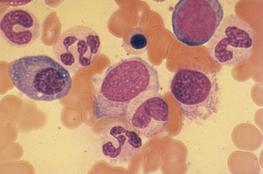
Blood cell production normally takes place in the bone marrow, and all the circulating cellular elements of the blood arise from the level of a pluripotent hematopoietic stem cell (Fig. 39-1). Hematopoietic stem cells have two important properties, extensive self-renewal and ability to differentiate and mature into red blood cells, granulocytes (neutrophils, eosinophils, basophils), monocytes, platelets, and lymphocytes (T cells, B cells, and NK cells). Little is known about the molecular mechanisms that control self-renewal, but this ability of stem cells is critical to the success of treatments such as bone marrow transplantation. More is known about the factors that control hematopoietic differentiation and maturation.


Hematopoietic stem cells are rare in number, estimated at approximately 1 in 1 × 10⁶ bone marrow cells, or 0.05% of all marrow cells. The fact that they exist at all is best defined by their ability to support bone marrow regrowth after stem cell transplantation or marrow recovery after severe marrow suppression from chemotherapy or radiation. Two major lineages arise from the pluripotent hematopoietic stem cell: the common myeloid progenitor cell and the common lymphoid progenitor cell. Lymphopoiesis then proceeds to the differentiation of T cells, B cells, natural killer (NK) cells, and some dendritic cells. The common myeloid progenitor gives rise to red cells, granulocytes of all types, monocytes and macrophages (and some dendritic cells), and platelets.


The regulation of the terminal stages of hematopoietic differentiation and maturation is under the control of a variety of hormones and cytokines. Red cell production is under the control of erythropoietin (EPO), a glycoprotein hormone secreted by the kidney in response to hypoxia or anemia. Neutrophil production is under the control of granulocyte colony-stimulating factor (G-CSF). Eosinophil production is under the control of interleukin-5 (IL-5). Monocyte and macrophage production is under the control of macrophage colony-stimulating factor (M-CSF) and, to a lesser extent, granulocyte-macrophage colony-stimulating factor (GM-CSF). Platelet production is under the control of thrombopoietin (TPO), produced in the liver. TPO also stimulates proliferation and release of hematopoietic stem cells. Of these various growth factors, EPO and G-CSF have important uses clinically; TPO is under clinical development. The number of cellular elements in the blood is tightly controlled by these regulatory hormones. Table 39-1 shows the normal ranges for the various cellular elements of the blood in adults.


Table 39-1 Cellular Elements of the Blood (Adult)


Red Blood Cells (RBCs) Men Women
Hematocrit (%) 46.0 ±4.0 40.0 ±4.0
Hemoglobin (g/dL) 15.7 ±1.7 13.8 ±1.5
RBC count (×106/μL) 5.3 ±0.7 4.6 ±0.5
Reticulocytes (%) 1.6 ±0.5 1.4 ±0.5
Mean corpuscular volume (fl) 88.0 ±8.0  
Mean cell hemoglobin (pg/RBC) 30.4 ±2.8  
Mean cell hemoglobin concentration (g/dL of RBC) 34.4 ±1.1  
Red cell volume distribution width (RDW) (%) 13.1 ±1.4  
Platelets (/μL) 150,000-300,000  
White Blood Cells Range (/μL) Range (%)
All white blood cells 4300-10,000 100
Total neutrophils 2000-7000 20-70
Segmented neutrophils 1500-6000 15-60
Band neutrophils 500-1000 5-10
Lymphocytes 1500-4000 15-40
Monocytes 100-900 1-9
Eosinophils 100-700 1-7
Basophils 0-150 0-1.5


Disorders of Red Blood Cells


Key Points


The major role of the red blood cell (RBC, erythrocyte) is to transport oxygen from the lungs to the tissues and organs in the body. This is accomplished by the reversible binding of oxygen to the heme moiety of the hemoglobin molecule. Hemoglobin (Hb) is the most prominent RBC protein. The functional ability of the Hb molecule is determined by primary amino acid structure, pH (Bohr effect), temperature, and intracellular concentration of 2,3-biphosphoglycerate (2,3-BPG). Abnormalities of Hb function most frequently result from mutations (amino acid substitutions) in one or more of the globin genes. These mutations, such as the mutation that causes sickle cell disease, can alter the stability of the Hb molecule or its oxygen-binding properties. Many of these mutations result in tissue hypoxia because the instability of the molecule results in the early death of the RBC in circulation (hemolysis). Other Hb mutations can affect oxygen binding and release or can produce free-globin chains that may bind to the inner RBC membrane, resulting in membrane instability (Heinz bodies). Hemoglobin is also recognized as the major “sink” of nitric oxide (NO) in the blood. This has important implications for the control of vascular tone in the microcirculation.


Under normal conditions, regulation of erythropoiesis can be viewed as a positive control loop. Red cell production is regulated by EPO. Production of EPO occurs primarily in the kidney, although a small amount is produced by the liver; this is a holdover from fetal life, when the liver was the major source of EPO production. EPO stimulates RBC production by binding to specific receptors on the surface of erythroid progenitor cells in the marrow. This activates a number of cell division steps that results in approximately 16 to 32 mature RBCs for each progenitor cell stimulated. These new RBCs leave the bone marrow and circulate for about 120 days as mature cells. The circulating red blood cells make up the RBC mass.


The RBC mass represents the oxygen-carrying capacity of the body. As RBCs traverse the circulation, the cells provide oxygen for all metabolic functions. In the kidney, oxygen availability is sensed by specialized peritubular capillary lining cells. The sensing mechanism is mediated by hypoxia-inducible factor 1α (HIF-1α). The adequacy of the oxygen supply depends not only on RBC mass, but also on normal lung function, a normal circulatory system that allows RBCs to be delivered effectively to all parts of the body, an adequate supply of EPO to stimulate RBC production, and a normally functioning bone marrow. Abnormalities in any of these factors can affect RBC production and result in anemia (too few RBCs) or erythrocytosis (too many RBCs).


Anemia


Key Points


Definition


Anemia is best defined as a reduced RBC mass resulting in decreased oxygen-carrying capacity of the blood. Table 39-1 lists normal values for hemoglobin and hematocrit (Hct); values below the lower limits of normal for age and gender indicate only 25% likelihood that the person is not anemic. Previous Hb and Hct values for a given patient are useful comparisons when interpreting currently determined levels. Both pregnant women and long-distance athletes may have increases in plasma volume so that the Hct or the Hb value, or both, fall artificially below normal ranges. They still have a normal oxygen-carrying capacity with a normal RBC mass and should not be considered anemic. Conversely, a dehydrated patient admitted to the hospital may have Hb or Hct that appears normal because of plasma volume contraction. Correction of their volume-depleted state will allow appropriate determination of their true RBC oxygen-carrying capacity. When Hct or Hb is abnormal, further investigation is indicated, whether or not the subject appears clinically well.


Prevalence


Mean hemoglobin concentration for all U.S. males older than 1 year is 14.67 g/dL, and for females older than 1 year, 13.19 g/dL (NHANES-III; NCHS, 2005b). These values vary by ethnic group; non-Hispanic white males had mean Hb of 14.05 g/dL, versus 13.87 g/dL for Mexican Americans and 13.14 g/dL for African Americans. Hb values for African American men and women are 1 g/dL lower than their white counterparts. The median Hb concentration was the same for both genders in all race and ethnic groups younger than 9 years. Mean Hb level tends to peak at 15.4 g/dL between ages 20 and 29 years and decreases to 14.36 g/dL by age 70. Anemia is defined as Hb level more than 2 standard deviations (SD) from the mean for age and race for the entire population.


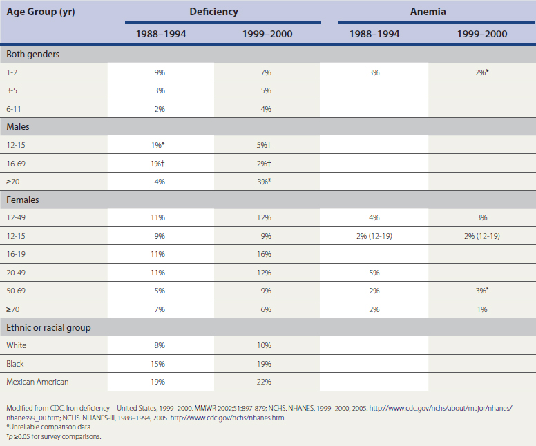
The most prevalent cause of anemia in the United States is iron deficiency. Table 39-2 categorizes the prevalence of iron deficiency and iron deficiency anemia (NHANES-III, 1988–1994, 1999–2000; NCHS, 2005a, 2005b). The prevalence of iron deficiency is 9% and of iron deficiency anemia 3%, in both genders age 1 to 2 years. The prevalence for children age 3 to 5 years is 3% for iron deficiency and less than 1% for iron deficiency anemia; those 6 to 11 years old have a 2% prevalence of iron deficiency and less than 1% for iron deficiency anemia. The prevalence of iron deficiency in the nonblack population is 1% lower than the prevalence in all races combined. Women have a much higher prevalence; those age 12 to 15 have 9% prevalence of iron deficiency and 2% for iron deficiency anemia; those age 16 to 19 have 11% and 3% prevalence; women 20 to 49 years, 11% and 5%; and those 50 to 69 years, 5% and 2%, respectively. In women over age 70, prevalence of iron deficiency rises to 7% and iron deficiency anemia to 2%. Males in general have less than a 1% prevalence of iron deficiency or iron deficiency anemia until age 50, when the prevalence of iron deficiency rises to 2%. In those older than 70, prevalence of iron deficiency is 4% and iron deficiency anemia 2%.


In a comparative study (Netherlands, Japan, Poland, United States) of diagnostic encounters, blood disorders comprised 1% of all encounters, and iron deficiency was the 27th most frequent diagnosis seen in all four countries (Okkes et al., 2002). Anemia is the most frequent hematologic disorder seen in family medicine and occurs often enough that knowledge of RBC function, classification of RBC disorders, evaluation of laboratory data, and treatment of common anemias constitute an important part of the knowledge base of the competent family physician.


Clinical Features


Anemia is most often recognized by abnormal screening laboratory test results. Much less frequently, patients will present to their family physician with previously unrecognized anemia, complaining of fatigue, loss of stamina, shortness of breath, and rapid heart rate (particularly with physical exercise). In younger patients, if the anemia comes on gradually, several compensatory mechanisms help maintain tissue oxygenation. These include peripheral vasodilation, increased cardiac output, a change in the oxygen-hemoglobin dissociation curve that facilitates oxygen unloading in the tissues, and shunting of blood away from circulation-rich organs (e.g., gut, skin, kidney) to critical organs (e.g., heart, brain).


Physicians must recognize that the signs and symptoms of anemia will be determined in part by the acuteness of onset. Acute anemia is almost always caused by blood loss or hemolysis. If blood loss is mild, enhanced oxygen delivery is achieved through changes in the oxyhemoglobin dissociation curve and in hemodynamics. With acute blood loss, however, the changes in blood volume dominate the clinical picture, and Hct and Hb levels do not reflect the volume of blood lost for at least 48 hours. Signs of vascular instability appear with acute blood losses of 10% to 15% of the total blood volume. In such patients, management issues are related not to the anemia, but to hypotension and decreased organ perfusion. When more than 30% of the blood volume is lost suddenly, patients are unable to compensate with the usual mechanisms of vascular contraction and changes in regional or organ blood flow. The patient prefers to remain lying flat and will show postural hypotension and tachycardia if placed in an upright position. If the volume of blood loss is more than 40% (>2 L in average-sized adult), signs of shock are prominent, including confusion, air hunger, sweating, hypotension, and tachycardia. These patients have significant deficits in vital organ perfusion and require immediate volume replacement.


For the family physician, certain disorders are associated more often with anemia. These include chronic inflammatory states (e.g., infection, rheumatoid arthritis, cancer) associated with mild to moderate anemia, whereas lymphoproliferative disorders (e.g., chronic lymphocytic leukemia, other B-cell neoplasms) may be associated with immune-mediated hemolysis.


Laboratory Evaluation


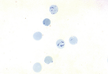
Key laboratory tests in the office evaluation of the anemic patient include complete blood count (CBC), reticulocyte count, and iron studies (Box 39-1). The laboratory evaluation of anemia is designed primarily to determine effective response of the bone marrow to the anemia stimulus and to detect any disturbance in iron metabolism; this information allows physiologic classification of the most common anemias. The evaluation of the marrow’s response to anemia is best approximated through corrected reticulocyte count (Fig. 39-2), which provides information about the number of newly released RBCs in circulation. Normally, a newly released RBC can be seen as a reticulocyte for about 24 hours. The reticulum of a reticulocyte is made up of residual ribosomal ribonucleic acid (RNA); the cell is somewhat larger and appears bluer than mature RBCs on a Wright-Giemsa–stained peripheral blood smear.


First, however, the raw reticulocyte percentage must be corrected if it is to reflect the marrow production index. The first correction of the reticulocyte count is for dilution, as shown by the following equation for determining reticulocyte index (RI):


image


The normal reticulocyte count is 1% to 2% and the normal RI is 1.0. This correction is not necessary if the laboratory reports reticulocyte count as an absolute number, normally about 40,000 to 50,000 cells/μL.


A further correction of the reticulocyte count is necessary if there is evidence from the peripheral blood smear that reticulocytes are being released prematurely from the bone marrow (shift cells or shift reticulocytes). Under these circumstances, reticulocytes live longer than the usual 24 hours in circulation, and thus the uncorrected reticulocyte count will overestimate the rate of new cell production. The second correction is shown by the equation for determining the marrow production index: MPI = RI/2. The normal MPI value is 1.0. The RI is divided by a factor of 2 to account for the prolonged reticulocyte life span in the circulation.


For example, if the reticulocyte count is 15% and Hct is 15%, there is evidence of shift reticulocytes on the peripheral smear. The MPI can be calculated as follows:


image


Also critical to understanding the pathophysiology of most anemias is to characterize the availability of iron for hemoglobin synthesis. This is done by measuring the serum iron level, total iron-binding capacity (TIBC), and serum ferritin level. Transferrin saturation percent is the proportion of iron binding sites occupied by iron atoms, reflecting the amount of iron immediately available for Hb synthesis, and is very labile. Serum ferritin is an indirect reflection of the body’s total iron stores and is more stable. These values provide information about the two most common forms of anemia seen in the hospital or in the family physician’s office—iron deficiency anemia and the anemia of chronic inflammation.


Physiologic Classification


The physiologic classification of anemia is based on response of the bone marrow. The three major categories are hypoproliferative anemia, maturation disorders (ineffective erythropoiesis), and hemolytic-hemorrhagic anemia (Box 39-2).


Hypoproliferative Anemia


In hypoproliferative anemias the response of the bone marrow is impaired by one of three general mechanisms. The first is marrow damage, which results from an injury to the bone marrow and makes it impossible for the marrow to respond to adequate EPO stimulation. Aplastic anemia is a classic example, but chemotherapy-induced marrow aplasia is more common (Box 39-3). The physiologic hallmark of marrow damage is a low RPI (<2 at Hb >10 g/dL; <2.5 at Hb of 7-10 g/dL). EPO is typically elevated, with evidence of premature release of reticulocytes from the bone marrow (shift reticulocytes, including nucleated RBCs in some cases). Despite EPO stimulation, however, the bone marrow is unable to proliferate. Bone marrow examination may reveal an empty or hypocellular marrow or one replaced by tumor cells or fibrosis. Treating these patients with recombinant human EPO is rarely useful.


A second mechanism of hypoproliferation is inflammation, which is the most common form of anemia seen in hospitalized patients. Often referred to as the “anemia of chronic disease” (ACD), it has more recently been termed the anemia of inflammation (AI) (Weiss and Goodnough, 2005). The anemia associated with inflammatory states is complex and involves altered iron homeostasis as well as decreased EPO production and an impaired response of erythroid progenitor cells in the marrow to EPO. Inflammation comes in many forms in addition to acute and chronic infection. Also associated with changes consistent with AI is tissue damage, as caused by necrosis, surgery, myocardial infarction, and other tissue injury.


The major effect of AI is on iron metabolism. With inflammation, iron absorption from the gut is blocked, as is iron release to transferrin from reticuloendothelial stores. The net effect is a decrease in the serum iron level and in transferrin saturation. This results in an inadequate supply of iron to the erythroid marrow for hemoglobin synthesis. Over time, the clinical picture (e.g., microcytic, hypochromic RBCs) may come to resemble true iron deficiency anemia, despite the fact that iron stores in the body are normal or increased. The mediator of these alterations in iron metabolism is hepcidin, a small molecule made in the liver that is critical to iron homeostasis and that is upregulated in the presence of inflammation. Hepcidin interferes with the cellular iron export protein ferroportin. The ferroportin pathway is the mechanism by which iron absorbed from the diet is shunted through the gut and reticuloendothelial cells and released to circulating transferrin. Iron regulatory proteins (IRP-1 and IRP-2) also play a role in balancing iron storage as well as circulating iron. Although there are many similarities to iron deficiency, providing iron in this setting is generally ineffective. Recombinant human EPO (epoetin) may stimulate RBC production, but the approach to treatment is primarily to identify and reverse, if possible, the inflammatory trigger.


In addition to changes in iron homeostasis, inflammation results in the release of proinflammatory cytokines such as IL-1, tumor necrosis factor alpha (TNF-α) and interferon γ (IFN-γ). These cytokines have many effects on erythropoiesis, such as decreasing EPO production and blunting the response of erythroid progenitors in the marrow to EPO. All these changes result in marrow hypoproliferation and an inadequate marrow response to anemia.


The third major category of hypoproliferative anemia is that associated with inadequate EPO production, which results in understimulation of the marrow. Typically, this is seen in patients with chronic renal insufficiency whose diseased kidney cannot produce EPO despite often profound anemia. The advent of successful renal replacement therapy (peritoneal or hemodialysis) has resulted in an increasingly large population of severely and chronically anemic patients. For these patients, epoetin therapy has been lifesaving in terms of quality of life and overall health.


In the family physician’s office, the question often is how much anemia can be ascribed to mild or moderate renal insufficiency. There is no easy answer, but generally, if the creatinine level is higher than 2 mg/dL with no other obvious or reversible cause for the anemia (blood loss, hemolysis after appropriate testing), it is reasonable to ascribe the anemia to renal insufficiency.


Another pathologic process resulting in inadequate EPO stimulation is hypometabolism, particularly hypothyroidism. The anemia may reflect a reduced need for oxygen-carrying capacity because of the reduced metabolic load resulting from the thyroid hormone deficiency.


Mild iron deficiency anemia also is associated with a hypoproliferative marrow response. Iron deficiency, with mild to moderate anemia, impairs the erythroid marrow response. If the anemia is mild, circulating RBCs are normocytic or slightly microcytic, and the red cell distribution width (RDW) index is normal (Fig. 39-3). The serum iron is usually low, transferrin saturation less than 15%, and serum ferritin less than 15 ng/mL.
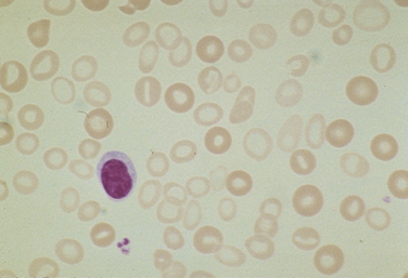


Table 39-3 compares anemia of inflammation and classic iron deficiency anemia. The major difference is the serum ferritin level, which is typically normal or increased with AI and characteristically low with true iron deficiency. Making this distinction is important because the mechanisms that lead to inflammation or iron deficiency are generally distinct, as is the approach to treatment.


Table 39-3 Comparison of Anemia of Inflammation and Iron Deficiency Anemia


Iron Deficiency Anemia of Inflammation
Low serum iron level Low serum iron level
Elevated TIBC TIBC normal or reduced
Transferrin saturation low (<15%) Transferrin saturation low (15%-20%)
Serum ferritin level low (<15 ng/mL) Serum ferritin level normal or elevated
Microcytic, hypochromic RBCs Normocytic to microcytic RBCs
RBC protoporphyrin level elevated RBC protoporphyrin level elevated

TIBC, Total iron-binding capacity; RBCs, red blood cells.


Maturation Disorders


Maturation disorders are characterized by adequate EPO stimulation and erythroid marrow hyperplasia, but in the absence of a sufficiently increased reticulocyte production index. Under these conditions, premature cell death (apoptosis) takes place in the marrow, and a mismatch occurs between degree of erythroid hyperplasia on bone marrow aspiration or biopsy and the reticulocyte (effective) production index. Consequently, RBC production in such patients is considered ineffective and can involve nuclear or cytoplasmic maturation defects.


Nuclear maturation defects result in a megaloblastic bone marrow and are typical in patients with severe folate or vitamin B12 deficiency. The RBCs are macrocytic, and the reticulocyte production index is normal or slightly above normal (Fig. 39-4). Examining the bone marrow of patients with vitamin B12 or folate deficiency reveals increased erythroid marrow precursors and loosening of nuclear chromatin, as well as more cells with nuclear degeneration (karyorrhexis and karyolysis). These are the features of apoptosis.
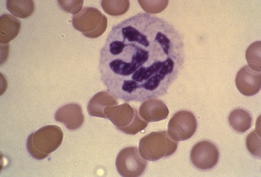


Because of the degree of RBC destruction in the bone marrow, serum bilirubin may be elevated and haptoglobin decreased. It is important to distinguish between vitamin B12 and folate deficiency because the pathogenesis is different and the treatment must be specific. Patients who present with folate deficiency and alcoholic neuropathy pose a particularly difficult diagnostic challenge; the neuropathy of vitamin B12 deficiency should not be treated inappropriately with folic acid, because the anemia may be partly corrected with folic acid, but the neuropathy associated with vitamin B12 deficiency will progress. This is rarely seen at present. The neurologic symptoms may precede the anemia, so it is important to screen older adults with unexplained memory loss for vitamin B12 deficiency.


The diagnosis of these disorders is relatively straightforward and can be made with laboratory testing for serum vitamin B12 and folate levels. RBC levels of folate may also be useful, particularly in a folate-deficient patient receiving diet therapy or an undernourished alcoholic patient being fed. Alcohol inhibits the entry of folic acid, decreasing serum folic acid levels. Thus, folic acid deficiency may result from both dietary insufficiency and inhibition of folic acid release in patients with chronic alcohol intake. Folic acid deficiency has greatly decreased since 1998, when most grains began to be fortified with folic acid in the United States.


Treatment of vitamin B12 or folic acid deficiency is simply replacement with the appropriate vitamin. In patients who appear with severe megaloblastic anemia and who need treatment immediately, it is prudent to obtain blood samples, both whole blood and serum, for later diagnostic tests and then to treat the patient with both vitamins. This ensures that the anemia and central nervous system (CNS) manifestations of potential vitamin B12 deficiency are adequately treated.


The Schilling test is useful to specify the defect leading to B12 deficiency. This involves a small, oral dose of radiolabeled vitamin B12 along with a large, parenteral flushing dose of B12. The B12 absorbed from the diet is excreted in the urine because there are no transport binding sites in the circulation. This first stage of the absorption test indicates if the patient can absorb vitamin B12. The second stage is similar to the first, except the oral dose of B12 is given with intrinsic factor (IF). Theoretically, the addition of IF should correct the absorption defect associated with pernicious anemia. If the absorption defect is caused by disease of the terminal ileum, both stages of the Schilling test will be positive. A host of surrogate markers for pernicious anemia include measurement of IF or anti–parietal cell antibodies. However, these markers are positive in an increasing percentage of patients as they grow older, and consequently cannot be considered definitive for the diagnosis.


Another cause of a macrocytic anemia associated with ineffective erythropoiesis is myelodysplasia. The myelodysplastic syndromes are primary bone marrow neoplasms, and the macrocytosis does not respond to vitamin replacement therapy. This is now an increasingly frequent diagnosis, particularly because it is a more common diagnosis in older adults and the population is aging.


Transient megaloblastic and macrocytic anemia may be seen with certain types of anticancer drugs that interfere directly with DNA synthesis and cell division. Common drugs in this category include hydroxyurea and thymidine inhibitors. Family physicians also see patients with mild anemia and macrocytosis who have a history of excessive alcoholic intake, with or without a mild to moderate degree of liver disease. The macrocytosis in these cases has two probable causes: spurious macrocytosis caused by lipid loading of the RBC membrane, and macrocytosis caused by intermittent folate deficiency associated with bouts of high alcohol consumption in the absence of other caloric intake.


Iron Deficiency

In addition to nuclear maturation defects that result in macrocytic anemia, cytoplasmic maturation defects such as severe iron deficiency occur. Extreme iron deficiency and severe anemia may result in a pattern of ineffective RBC production. With mild anemia, iron deficiency limits erythroid proliferation, and the anemia appears to be hypoproliferative. However, as anemia becomes more profound and EPO stimulation of the marrow increases, the marrow becomes more ineffective in appearance. In either case, laboratory test results indicating low serum iron, low transferrin saturation, and extremely low ferritin are diagnostic.


Once the diagnosis of iron deficiency is definitive, the family physician needs to consider the cause. Women in their childbearing years typically have marginal iron stores, and even being an occasional blood donor may result in mild anemia, with iron deficiency. The case is different in an adult male or postmenopausal woman. Unless there is a clear explanation, gastrointestinal (GI) blood loss should be the prime suspect and must carefully be ruled out.


Treatment of iron deficiency is not always straightforward. The goal is to remedy the hemoglobin deficit and replace iron stores. Many oral iron preparations are available, but ferrous sulfate and ferrous gluconate are most often used and inexpensive. The best regimen is to give 3 iron tablets daily; this provides about 150 mg of elemental iron daily. If the patient is compliant and absorption normal, reticulocytosis will occur in a week and Hb level will increase at least 1 g/dL within 2 weeks. Microcytosis may take up to 4 months to resolve. The iron is best taken on an empty stomach because certain foods interfere with iron absorption. However, 15% to 20% of patients will have significant gastric upset with oral iron and may not be compliant. If poor absorption or noncompliance is of concern, parenteral iron may be given. Several preparations are available, including iron sucrose and iron gluconate intravenously (IV) at 125 to 250 mg/day, and have a good safety profile compared with iron dextran. In treating iron deficiency, the target for therapy is not just to correct the anemia, but also to provide some degree of iron stores, so it is recommended that iron treatment be continued for 2 to 3 months after a normal Hb level is reached.


Various preparations of ferrous salts are equally tolerated and effective for the treatment of iron deficiency anemia. Controlled-release (CR) iron formulations cause fewer GI side effects than non-CR salt preparations, but discontinuation rates are similar. Ferrous salts are the treatment of choice for iron deficiency anemia (McDiarmid and Johnson, 2002).


Hemochromatosis in hereditary form has a homozygous incidence of 0.44% and a 10% heterozygous incidence, all in populations of white European descent. Elevated transferrin saturation levels (>45%-50%) and elevated ferritin levels (>300 mg/L) are markers of increased iron stores that may indicate the need for further evaluation.


Hemolytic-Hemorrhagic Anemia


Hemolytic-hemorrhagic anemia (HHA) is diagnosed in patients with persistent anemia or decreasing Hb and Hct levels despite what appears to be an adequate bone marrow erythropoietic response. For patients with chronic hemolytic disease, this means a marrow production index of 3. In patients with hemorrhagic disease, this level of production may not be reached quickly because of the ongoing iron loss, and production indices about 2 to 2.5 times normal are more common. In the latter case, however, blood loss dominates the clinical picture unless the loss is internal.


In a discussion of the hemolytic anemias, which can be complex to diagnose, it is helpful to consider the pathophysiology of disease and how to categorize the hemolytic process. One should first consider whether the hemolysis appears to be intravascular or extravascular.


Intravascular Hemolysis

Intravascular hemolysis is associated with the rupture of erythrocytes and dispersion of their contents into the plasma. This results in free hemoglobin in the plasma and, if sufficient RBC destruction takes place, there is Hb spillover into the urine (hemoglobinuria). The primary Hb-binding protein in the plasma is haptoglobin, but in the presence of intravascular hemolysis, free haptoglobin becomes undetectable. This is a useful test only if the results are negative because ineffective RBC production, associated with substantial destruction of RBCs in the bone marrow (or even primarily extravascular hemolysis), will result in the release of sufficient Hb to reduce circulating haptoglobin levels.


Intravascular hemolysis can be life threatening if acute and can have several causes. For example, RBC membrane damage and intravascular hemolysis can result from burns or exposure to certain toxins that target the RBC membrane, such as Clostridium perfringens. Hypotonic lysis is rarely seen but could occur because of the IV infusion of free water. Immune-mediated lysis of RBCs can occur when mismatched transfusions are given. With ABO incompatibility, the operative mechanism is immunoglobulin M (IgM) antibodies that fix complement to the RBC surface and cause rapid lysis.


Mechanical fragmentation is probably the most common form of intravascular hemolysis seen in North America and is associated with microvascular diseases such as thrombotic thrombocytopenic purpura, hemolytic uremic syndrome (HUS; Fig. 39-5), defective mechanical heart valves, and disseminated intravascular coagulation. Acute attacks of malaria are also associated with intravascular hemolysis. Paroxysmal nocturnal hemoglobinuria (PNH) is an unusual form of intravascular hemolysis caused by an acquired X-linked defect in the hematopoietic stem cell. Patients with PNH have varying degrees of hemolysis throughout the day, and in crisis, this can be severe.
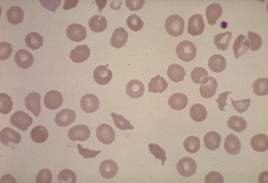


An inherited condition that predisposes patients to intravascular hemolysis is the Mediterranean form of glucose-6-phosphate dehydrogenase (G6PD) deficiency. G6PD deficiency is the most common inborn error of RBC metabolism worldwide, affecting almost 0.5 billion people. Because it is an X-linked genetic disorder, it is most severe in men. G6PD protects RBCs from oxidative stressors such as superoxide anions and hydrogen peroxide. Oxidative products in RBCs are normally neutralized by reduced glutathione, which is generated by glutathione reductase. This pathway is part of the hexose monophosphate shunt pathway and requires G6PD for proper functioning. The Mediterranean abnormality of G6PD deficiency is associated with moderate chronic hemolysis, but this may become overwhelming and even fatal when the RBCs are exposed to oxidative stress. Any oxidative stress in these patients may have severe clinical consequences that could require transfusion or exchange transfusion as part of therapy.


The more common variety of G6PD deficiency in North America is G6PDA-, which affects approximately 10% of the African American population. G6PDA- is an enzyme with reduced stability and catalytic activity that become more prominent as the red cell ages. In these people, hemolytic events typically involve older RBCs and are therefore benign and relatively self-limited. The accumulation of oxidized glutathione reacts with hemoglobin and causes precipitation of Hb into Heinz bodies, resulting in hemolytic disease. The hemolytic process can be enhanced by infection, surgical stress, diabetic acidosis, and certain medications, such as the antimalarials primaquine and quinacrine.


Sickle Cell Anemia


Sickle cell anemia is an inherited autosomal condition in which glutamic acid in the sixth position on the β-globin chain is replaced by a valine (Glu6Val). This results in hemoglobin SS in the homozygous state. Sickle cell trait, or hemoglobin AS, is found in 8% to 10% of African Americans in the United States, and sickle cell anemia occurs in about 1 in 400, or about 70,000 individuals. The gene for HbS is prevalent in sub-Saharan Africa. Persons of Mediterranean descent from India or Saudi Arabia have varying but somewhat lower percentages of the carrier state for HbSS. Sickle cell anemia occurs worldwide but predominates in Mediterranean, Saudi Arabian, and Indian populations. This distribution appears to be associated with independent mutations in these regions. People with hemoglobin AS or SS are somewhat protected against malaria because their erythrocytes are resistant to invasion by malarial parasites. If the parasite infects the cell, the rate of sickling increases, causing the cell with the parasite to be removed from the circulation more rapidly.


The pathophysiology of the sickling process is related to the oxygenation of the molecule. When deoxygenated, HbSS tends to polymerize into long, tubelike fibrils. This results in an elongated, rigid cell subject to trapping in the microcirculation, resulting in vaso-occlusive crisis. Hypoxemia, acidosis, dehydration of the RBC, hyperosmolality of the renal medulla, and viral infections can play a role in triggering or accentuating the sickling process. Sickled RBCs are also more adhesive to endothelial cells, activate endothelial cells, and contribute to the interaction between neutrophils and activated endothelium in the microvasculature. When the conditions that cause sickling are corrected, the sickle cell may return to a more normal shape and function. However, some sickled RBCs are irreversibly changed, indicating that the membrane cytoskeleton has been damaged. Irreversibly sickled cells are generally removed in the sinusoidal RES networks, but approximately one-third may be hemolyzed intravascularly. Some degree of anemia has a protective effect in vaso-occlusive crises by helping reduce blood viscosity and deoxygenation, thereby reducing the likelihood of polymerization.


During vaso-occlusive crises, the microvascular capillaries, capillary bed, and veins become occluded. Contributing to this locally are low pH, prolonged capillary transit time, and infection. Increased numbers of sickled RBCs occlude the vessels, which leads to painful ischemia, infarction, and reduced organ function over time. Areas particularly affected include the portal circulation, in which oxygen tension is low; the kidney, in which the renal medulla is hyperosmolar and dehydrates RBCs, increasing mean corpuscular hemoglobin concentration (MCHC); the lungs; and the brain. It is not clear why some patients have severe, frequent episodes and others do not. Avascular necrosis of the bone marrow is usually the cause of severe pain.


Clinical Diagnosis


A patient with sickle cell disease usually has chronic hemolysis that results in a moderate anemia, intermittent jaundice, and a marrow with a thinned, bony cortex. Vaso-occlusive episodes and the appearance of Howell-Jolly bodies occur by the time the patient becomes an adult. The presence of Howell-Jolly bodies indicates that the patient has undergone an autoinfarction of the spleen.


The diagnosis of sickle cell disease is easily made by finding sickled cells on the peripheral smear, preparing a sickle cell sample, and carrying out a screening hemoglobin electrophoresis, which will confirm the Hb type. For a sickle cell preparation, blood is mixed with 2% sodium metabisulfite, which produces sickling. The proportion of sickled cells is measured initially and then 1 hour later to make the diagnosis. The “gold standard” diagnosis is Hb electrophoresis, which shows the relative amounts of Hb forms.


Patients with long-standing sickle cell anemia may have pain syndromes related to bone marrow vaso-occlusive crises and aseptic necrosis of the femoral head. Bilirubin gallbladder stones occur in 40% to 60% of patients. Hepatic and cardiac complications may also develop. Other associated problems include vascular occlusion in the pulmonary bed, leading to acute chest syndrome. Breakdown of the skin over bony prominences in the lower extremities is common and may lead to chronic, poorly healing ulcers. Papillary necrosis of the kidney may occur and, with age, renal function may decline. In the eye, conditions such as hemorrhage and neovascularization may occur. Patients with sickle cell anemia are particularly susceptible to Salmonella infections because of decreased complement activation, and asplenic patients are also subject to an increased likelihood of infection from encapsulated organisms such as Pneumococcus. Patients with sickle cell anemia should be given Pneumovax vaccine to prevent streptococcal pneumonia and sepsis. Haemophilus influenzae infection is responsible for a high percentage of pneumonia cases in sickle cell anemia patients.


Neurologically, patients with sickle cell anemia are subject to cerebrovascular accident (CVA, stroke). Particularly disturbing, children with severe sickle cell anemia will have evidence of multiple small strokes on magnetic resonance imaging (MRI) or computed tomography (CT) of the head by age 10 years. This has been associated with learning impairments in affected children. Aggressive transfusion protocols have been tested to reduce the incidence of this complication, with some success. Thrombosis is more common in children, whereas hemorrhage is more common in adults, possibly because of the occlusion of small vessels in hypoxic situations. Also, sickled cells show increased attraction to the endothelium, causing proliferation of the endothelial intimal surface, which can contribute to vascular occlusion. There is a high rate of recurrent stroke within 3 years of the initial event. Parvovirus infection can also cause pure RBC aplasia in these patients and lead to a devastating exacerbation of the anemia.


Treatment


Patients with sickle cell anemia should have Hb and Hct maintained at moderately low level to protect against vaso-occlusive complications related to viscosity. Patients with a painful crisis should be put on bed rest, hydrated vigorously, and given oral analgesics. Nonsteroidal anti-inflammatory drugs (NSAIDs), antihistamines, and benzodiazepines may also be helpful. Mild alkalinization of the blood is recommended. Oxygen should be given by nasal cannula or mask to keep the hemoglobin well oxygenated. Parenteral narcotics are rarely necessary, especially if the patient is given large volumes of fluid, oxygen, and oral morphine (or similar preparation) for pain control. Avascular necrosis of the bone marrow causes severe pain, and the patient may require intravenous (IV) narcotics and hydration for pain control.


Treatment with drugs that increase the proportion of fetal hemoglobin (HbF) show promise. Mixed polymers of HbF and HbS do not have the same propensity to sickle as SS tetramers. Azacitidine (5-AZA) increases the proportion of HbF but has significant toxicity. Hydroxyurea increases HbF level, which often results in fewer painful episodes, hospital admissions, and transfusions. The goal is to raise HbF level, if possible, to 20% to 30% of the total hemoglobin. Mortality can be reduced by as much as 40%. Unfortunately, not all patients respond to these types of interventions. Because both are antitumor drugs, azacitidine and hydroxyurea have significant toxicity, such as tumor induction. Gene therapies are being investigated but are not yet successful. In severe situations, allogenic (sibling) bone marrow transplantation has been tried, resulting in an 86% event-free survival at 5 years after transplant (Bhetia and Walters, 2008).


Patients undergoing surgery with general anesthesia are at increased risk of hypoxemia, sickling, and sickle cell crisis. They should be transfused before surgery with packed RBCs to bring the HbA level to 50% or higher. This decreases the likelihood of sickle cell episodes.


Pregnancy increases the risk of vaso-occlusive crisis for the mother. There is a high incidence of fetal loss among women who have sickle cell anemia. Vaso-occlusive episodes in the placenta may result in lower-birth-weight infants or fetal death.


Acquired Forms of Hemolysis


The acquired forms of reticuloendothelial or extravascular hemolysis are dominated by patients with acquired immune-mediated hemolytic disease. Patients with autoimmune hemolytic anemia (AHA) can present with severe anemia; this is usually associated with another disease such as a collagen vascular disease (e.g., rheumatoid arthritis, severe systemic lupus erythematosus) or drugs, or it is idiopathic. AHA is uncommon but can be dramatic on presentation. About 50% of patients have no associated disease. The Coombs antiglobulin test, performed directly and indirectly determines the presence of antibody coating the patient’s RBCs or free antibody in the plasma capable of binding to RBCs in AHA diagnosis. The autoantibodies may fix complement or may target the red cell for phagocytosis by the RES.


Therapy of AHA depends on the severity and causative mechanism. If the AHA is drug-induced, it is often sufficient to stop the drug and wait for the antibodies to clear, although this may take some time. Drug-induced hemolytic disease is uncommon and requires a specialized laboratory to make a precise diagnosis. AHA associated with collagen vascular diseases, lymphomas, or Hodgkin’s disease may be controlled with treatment of the primary underlying condition. The mainstay of treatment of AHA is high-dose corticosteroid therapy, at least 1 mg/kg of prednisone daily; refractory cases may require up to 2 mg/kg/day. Patients who do not respond to steroid therapy may be candidates for splenectomy, because the spleen is the primary site of removal of antibody-coated RBCs. Other drugs used as second- and third-line therapies for their immunosuppressive effects include cyclophosphamide, chlorambucil, and intravenous immune globulin (IVIG).


A final mechanism of acquired extravascular hemolysis is hypersplenism. Typically, patients with hypersplenism have an enlarged spleen because of infiltrative disease of the spleen, or they have splenomegaly as a result of portal hypertension. Under these circumstances, the RBCs are trapped for an unusually long period in the splenic circulation. Splenectomy may be beneficial.


Polycythemia


Polycythemia (or erythrocytosis) is the overproduction of red blood cells (erythrocytes). The mechanism of action resulting in polycythemia may be primary or secondary. Primary polycythemia indicates that the disorder arises at the level of the hematopoietic stem cells (see later), whereas secondary polycythemia represents the overproduction of RBCs caused by the increased stimulation of the bone marrow by EPO.


Polycythemia is typically identified initially in the laboratory by Hb and Hct values that are substantially above normal, requiring an explanation. Hb values of 18 to 20 g/dL lie in a range in which it is unclear whether the elevated Hb is caused by increased RBC production or a decreased plasma volume. In the latter case, the elevated Hb and Hct levels are caused by a contraction of the plasma volume and not by an increase in the RBC mass, so-called spurious polycythemia (pseudopolycythemia), or Gaisbock’s syndrome, and may not warrant therapeutic intervention. Typically, Hb level above 20 g/dL is unequivocally associated with increased RBC mass.


In the evaluation of patients with polycythemia or suspected polycythemia, it is important to first establish that the RBC mass has increased. This is achieved through direct measurement of the red cell mass using isotope dilution techniques, and the test can be performed at most large hospitals. Once that issue has been resolved, and assuming that there is evidence of RBC overproduction, it is important to determine whether the polycythemia is primary or secondary by measuring the circulating EPO level. If elevated, causes of the increased EPO production to consider include heart or chronic pulmonary disease, in which there is desaturation of Hb as it leaves the lungs, or less common causes such as renal cysts, hepatic or cerebellar tumors, uterine leiomyoma, or impaired Hb function caused by heavy smoking, which results in elevated levels of carboxyhemoglobin, which is essentially inert as an oxygen transporter. Severe obstructive sleep apnea may cause enough desaturation to raise EPO levels, resulting in secondary polycythemia. Athletes using EPO, androgens, or blood doping may also present with secondary polycythemia.


Certain forms of polycythemia are inherited. These can be associated with mutations in the Hb molecule that increase Hb’s affinity for oxygen, making it more difficult to offload oxygen within the tissues. These are referred to as “high-affinity mutants,” and more than 20 have been identified. It is important to identify the various possibilities because different causative factors have different long-term consequences.


Primary polycythemia, or polycythemia vera (PV), is a disorder of the hematopoietic stem cell. It is acquired typically in midlife and thereafter and, when full-blown, is associated with an increase in the platelet count, white cell count, splenomegaly, and severe itching following hot showers or baths. Pruritus can be disabling and presumably results from degranulation of skin basophils. Unlike secondary forms of erythrocytosis, EPO level is in the normal or low-normal range.


Treatment of PV strives to reduce the RBC mass to normal. Because PV is usually associated with an increase in total blood volume, the goal of phlebotomy therapy should be Hb and Hct levels in the normal range. Eventually, repeated phlebotomies induce iron deficiency, which is a reasonable end point. For patients with more aggressive disease (e.g., a growing spleen, increasing white cell and platelet counts), hydroxyurea therapy is effective. The goal is to maintain blood counts as close to normal as possible, or at least in an acceptable range.


Myeloproliferative Disorders


Polycythemia vera is one of a group of conditions referred to as the myeloproliferative disorders. These include essential thrombocytosis (ET) and agnogenic myeloid metaplasia with myelofibrosis (AMM/MF), also known as idiopathic myelofibrosis. All these disorders are characterized by the overproduction of one or more cell types. In PV the clinical picture is dominated by the increased production of RBCs. In ET the clinical picture is dominated by increased numbers of platelets, which may reach values as high as 2 × 10⁶/μL. With AMM/MF the clinical picture is dominated by various cytopenias associated with marrow fibrosis and splenomegaly. Splenomegaly, in this case, likely represents extramedullary hematopoiesis.


Many features of these diseases overlap. In PV, the spent phase may resemble myelofibrosis. Some patients with ET will eventually manifest an increased RBC mass. The occasional overlapping and thus confusing nature of these disorders has been partially explained by the recent demonstration of a very high frequency of a point mutation in a signaling molecule on the EPO signaling pathway (Kralovics et al., 2005). In several reports, almost two thirds to three quarters of patients with PV had this mutation in JAK2, with lower frequencies seen in patients with ET and AMM/MF. The mutation results in a constitutively activated pathway and the overproduction of bone marrow cellular elements. It can be anticipated that this will become a useful molecular test in the differential diagnosis of these disorders and will almost certainly become a target for drug development.


In ET the peripheral smear reveals increased numbers of platelets of various sizes. Platelet counts may be high, in excess of 1 or 2 × 10⁶/μL. There may be an associated leukocytosis, and Hb level is typically normal. The decision to treat such patients is not always easy. Generally, patients with platelet count approaching 1 × 10⁶/μL are treated, even with no associated symptoms or signs. Asymptomatic young women with ET require special consideration; watchful waiting is probably sufficient, although hydroxyurea plus aspirin is the most effective approach for patients who require treatment.


Patients with AMM/MF tend to run an indolent course. Usually, over time, the spleen continues to enlarge, and peripheral RBC destruction increases. There also may be varying degrees of thrombocytopenia caused by impaired bone marrow production and increased splenic sequestration. In all the myeloproliferative disorders, platelet function may be abnormal. Treatment of AMM/MF is largely symptomatic. The anemia can be treated with transfusions, or epoetin therapy can be attempted, although no good clinical trials support these approaches. In advanced cases of myeloid metaplasia, the spleen may grow to such a size that it may require removal to stabilize peripheral blood counts. Proper management of all the myeloproliferative disorders is associated with extended survival.


Oct 3, 2016 | Posted by in MANUAL THERAPIST | Comments Off on Hematology

Full access? Get Clinical Tree

Get Clinical Tree app for offline access