86
Giant Cell Tumor of the Tendon Sheath (Benign)
Kevin D. Plancher
History and Clinical Presentation
A 40-year-old right hand dominant woman presented with a mass on her left index finger. The patient reports that she has no symptoms related to the mass; however, occasionally she notices pain, stiffness, and numbness. The patient has had the mass for ∼2 years and does not recall any specific trauma to the area. She would like the mass removed for cosmetic reasons and because it causes her some functional problems.
Physical Examination
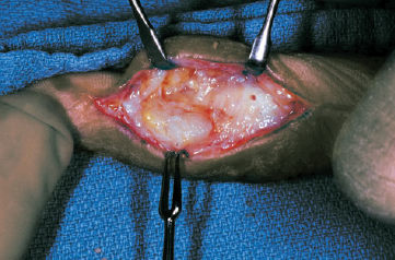
A mass is palpated on the volar ulnar aspect of the patient’s left index finger (Fig. 86–1). The mass is nontender, firm, and fixed. The skin over the mass is mobile. The mass is not transilluminating.
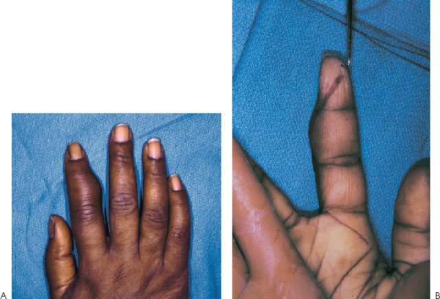
Figure 86–1. (A,B) Giant cell tumor of left index finger, ulnar aspect, with (A) dorsal and (B) volar views.
Diagnostic Studies
Standard radiographs show a soft tissue mass. Adjacent to the mass, cortical erosion, from the pressure effect of the mass, is seen. Bony erosion is not an uncommon finding with some giant cell tumors of the tendon sheath.
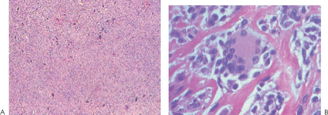
Histologic analysis reveals spindle cells, multinucleated giant cells, mononuclear cholesterol-laden histiocytes, fibroblasts, and round oval synovial cells arranged in clusters (Fig. 86–2).
Differential Diagnosis
Foreign body granuloma, necrobiotic granuloma
Fibroma of the tendon sheath
Infection
Ganglion cyst
Rheumatoid nodule
Epidermoid cyst
Lipoma
Giant cell tumor of the tendon sheath
Diagnosis
Giant Cell Tumor of the Tendon Sheath
Giant cell tumors of the tendon sheath are the second most common tumors of the hand, with simple ganglion cysts being the most common. These tumors were first characterized by Chassaignac in 1852. Giant cell tumors of the tendon sheath most commonly occur in patients in the third to fifth decades of life. The tumors are rare in young children and in the elderly. Giant cell tumors of the tendon sheath are often associated with degenerative joint disease.