Fig. 56.1
Plain x-ray. Giant cell granuloma producing a lytic lesion in the base of the distal phalanx
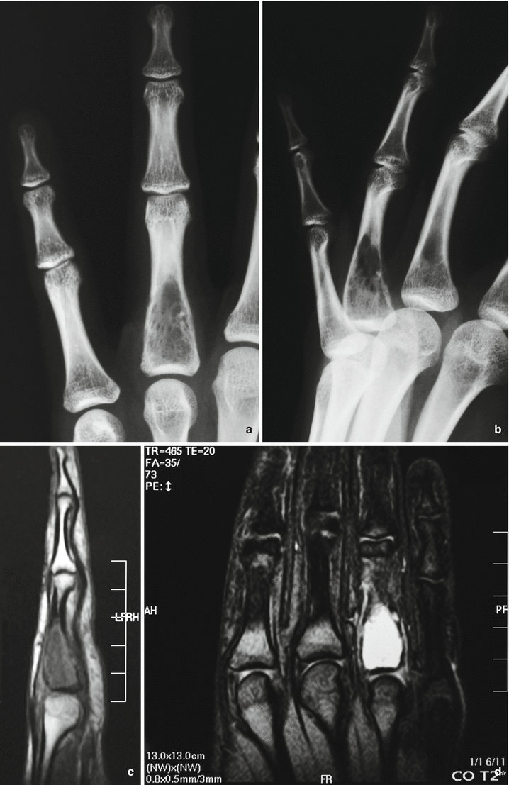
Fig. 56.2
(a, b) Anteroposterior and lateral x-ray of the hand. Proximal end of the first phalanx with a purely lytic well-marginated lesion. (c, d) T1- and T2-weighted MRI of the lesion, showing a hypointense and high-intense image
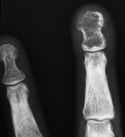
Fig. 56.3
X-ray. Giant cell granuloma in the third phalanx with erosion of the cortex
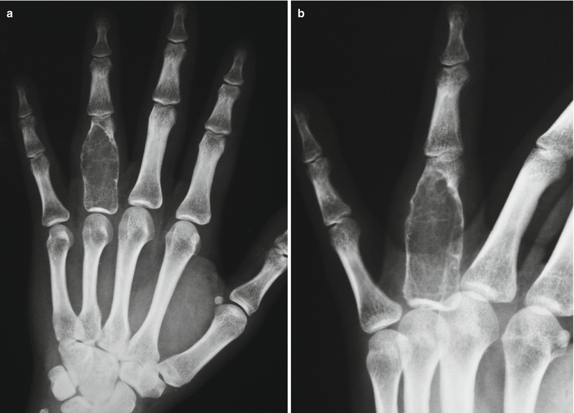
Fig. 56.4
(a, b) Anteroposterior and lateral view. Giant cell granuloma forming an expansile lesion in the first phalanx