General Principles of Infection

Chapter 20 General Principles of Infection


Etiology


Bone and joint infections pose a formidable challenge to the orthopaedic surgeon. The high success rate obtained with antibiotic therapy in most bacterial diseases has not been obtained in bone and joint infections because of the physiological and anatomical characteristics of bone. The overall surgical site infection rate has been estimated by the U.S. Centers for Disease Control and Prevention (CDC) to be 2.8% in the United States. Although bacteremia is common—estimated to occur 25% of the time after simple tooth brushings—other etiological factors must be present for an infection to occur. The mere presence of bacteria in bone whether from bacteremia or from direct inoculation is insufficient to produce osteomyelitis. Illness, malnutrition, and inadequacy of the immune system also can contribute to bone and joint infections. As in other parts of the body, bones and joints produce inflammatory and immune responses to infection. Osteomyelitis occurs when an adequate number of a sufficiently virulent organism overcomes the host’s natural defenses (inflammatory and immune responses) and establishes a focus of infection. Local skeletal factors also play a role in the development of infection. For example, the relative absence of phagocytic cells in the metaphyses of bones in children may explain why acute hematogenous osteomyelitis is more common in this location.


The peculiarity of an abscess in bone is that it is contained within a firm structure with little chance of tissue expansion. As infection progresses, purulent material works its way through the haversian system and Volkmann canals and lifts the periosteum off the surface of bone. The combination of pus in the medullary cavity and in the subperiosteal space causes necrosis of cortical bone. This necrotic cortical bone, known as a sequestrum, can continue to harbor bacteria despite antibiotic treatment. Antibiotics and inflammatory cells cannot adequately access this avascular area, resulting in failure of medical treatment of osteomyelitis.


Recognizing these unique characteristics of bone infections, the best course is prevention. The orthopaedic surgeon should evaluate the risk of infection in each patient by considering patient-dependent and surgeon-dependent factors. Patient-dependent factors include nutrition, immunological status, and infection at a remote site. Surgeon-dependent factors include prophylactic antibiotics, skin and wound care, operating environment, surgical technique, and treatment of impending infections, such as in open fractures. Simply stated, it is much easier to prevent an infection than it is to treat it.


Patient-Dependent Factors


Nutritional Status


A patient’s nutritional status and immunological response are important. If a patient is malnourished or immunocompromised and cannot mount a response to an infection, the effects of any treatment are diminished. Malnutrition adversely affects humoral and cell-mediated immunity, impairs neutrophil chemotaxis, diminishes bacterial clearance, and depresses neutrophil bactericidal function, the delivery of inflammatory cells to infectious foci, and serum complement components. Basal energy requirements of a traumatized or infected patient increase from 30% to 55% of normal. Fever of just 1°F above normal increases the body’s metabolic rate 13%. Nutritional status can be determined preoperatively by (1) anthropometric measurements (height, weight, triceps skin fold thickness, and arm muscle circumference), (2) measurement of serum proteins or cell types (lymphocytes), and (3) antibody reaction to certain antigens in skin testing. Nutritional support is recommended before elective surgery for patients with recent weight losses of more than 10 lb, serum albumin levels less than 3.4 g/dL, or lymphocyte counts of less than 1500 cells/mm3, which can be obtained from a routine compete blood cell count and SMA-24. With the use of serum albumin and transferrin levels, the formula that follows can be used to screen for patients who may need nutritional support: [(1.2 × serum albumin) + (0.013 × serum transferrin)] − 6.43. If the sum is 0 or a negative number, the patient is nutritionally depleted and is at high risk for sepsis. If nutritional support is needed, enteral therapy should be used when the gastrointestinal tract is functional; if not, hyperalimentation must be employed.


Immunological Status


To fight infection, the patient must mount inflammatory (white blood cell count) and immune (antibody) responses that initially stop the spread of infection and then, ideally, destroy the infecting organisms. The body’s main defense mechanisms are (1) neutrophil response, (2) humoral immunity, (3) cell-mediated immunity, and (4) reticuloendothelial cells. A deficiency in production or function of any of these predisposes the host to infection by specific groups of opportunistic pathogens. Deficiencies in the immune system may be acquired or may result from congenital abnormalities. Immunocompromised hosts are not susceptible to all opportunistic pathogens. The susceptibility to a microorganism depends on the specific defect in immunity. Abnormal neutrophils or humoral and cell-mediated immunities have been implicated in infections caused by encapsulated bacteria in infants and elderly patients, in the increased incidence of Pseudomonas infections in heroin addicts, and in Salmonella and Pneumococcus infections in patients with sickle cell anemia.


Diabetes, alcoholism, hematological malignancy, and cytotoxic therapy are common causes of neutrophil abnormalities. If the neutrophil count decreases to less than 55/mm3, infections caused by Staphylococcus aureus, gram-negative bacilli, Aspergillus organisms, and Candida organisms become a major threat.


Immunoglobulins and complement factors are two plasma proteins that play crucial roles in humoral immunity. Patients with hypogammaglobulinemia or who have had a splenectomy are at increased risk of infection caused by encapsulated bacteria, such as Streptococcus pneumoniae, Haemophilus influenzae, and Neisseria organisms. When a defect in a component of the complement cascade is present, S. aureus and gram-negative bacillus infections are common. Septic arthritis caused by unusual organisms such as Mycoplasma pneumoniae and Ureaplasma urealyticum has been reported and should be suspected in patients with hypogammaglobulinemia and culture-negative septic arthritis.


Cell-mediated immunity depends on an interaction between T lymphocytes and macrophages. Primary cell-mediated deficiencies are rare, but secondary cell-mediated deficiencies are common. Corticosteroid therapy, malnutrition, lymphoma, systemic lupus erythematosus, immunodeficiency in elderly patients, and autoimmune deficiency syndrome all can cause a secondary cell-mediated deficiency, which predisposes the host to fungal and mycobacterial infections and also infection with herpesvirus and Pneumocystis jiroveci.


Vaccinations also play a role in host response. The hepatitis B vaccine has reduced the incidence of hepatitis B virus, and the H. influenzae type B vaccine that is given to children has all but eliminated musculoskeletal infections caused by H. influenzae.


Surgeon-Dependent Factors


Skin Preparation


Wound contamination exists whenever the skin barrier is broken, but proper skin preparation decreases the contamination caused by bacteria present on the skin. Skin barriers also may decrease skin contamination during surgery. Although the skin can never be disinfected completely, the number of bacteria present can be reduced markedly before surgery. The skin and hair can be sterilized with alcohol, iodine, hexachlorophene, or chlorhexidine, but it is almost impossible to sterilize the hair follicles and sebaceous glands where bacteria normally reside and reproduce. Skin preparations have a limited effect on sebaceous glands and hair follicles because they do not penetrate an oily environment. Disinfectants that penetrate the oily environment are absorbed by the body and have potentially toxic side effects. Hexachlorophene has better penetration but also has neurotoxic side effects.


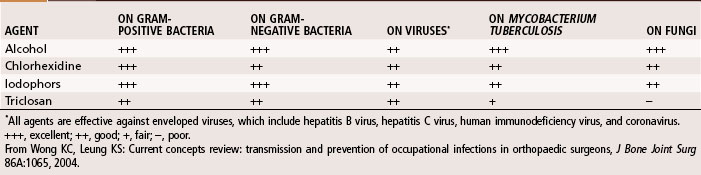
Hand washing is the most important procedure for prevention of nosocomial infections. Studies suggest that hand scrubbing for 2 minutes is as effective as traditional hand scrubbing for 5 minutes. The optimal duration of hand scrubbing has yet to be determined. Hand rubbing with an aqueous alcohol solution that is preceded by a 1-minute nonantiseptic hand washing for the first case of the day was found by Parienti et al. to be just as effective in prevention of surgical site infections as traditional hand scrubbing with antiseptic soap. The effectiveness of common antiseptics is summarized in Table 20-1.


Hair removal at the operative site is not recommended unless done in the operating room. Shaving the operative site the night before surgery can cause local trauma that produces a favorable environment for bacterial reproduction.


Prevention of infection transmission between the patient and the surgeon also includes proper surgical attire. Edlich et al. showed that a narrow glove gauntlet (cuff) significantly increased the security of the gown-glove interface. The U.S. Food and Drug Administration accepts a 2.5% failure rate of new unused sterile gloves. Glove perforation has been reported to occur in 48% of operations. Perforations usually occur approximately 40 minutes into the procedure, and as much as 83% of the time the surgeon is unaware of the perforation. Most frequently, the perforation occurs on the index finger or thumb of the nondominant hand. Double gloving reduces the exposure rate by as much as 87%. In addition, double gloving decreases the volume of blood on a solid needle (through a wipe clean pass mechanism from the outer glove) as much as 95%. A meta-analysis by Tanner and Parkinson found that double gloving decreased skin contamination, and the use of Biogel indicator gloves (Regent Medical, Norcross, GA) increased the awareness of glove perforation. When both gloves were compromised, however, the indicator gloves did not increase the awareness of a perforation. As long as the indicator glove was intact, perforation of the outer glove was promptly detected in 90%. Wearing an outer cloth glove over a latex glove significantly reduced the number of perforations to the innermost latex glove. When a liner glove was used between two latex gloves, the perforation rate of the innermost glove decreased. No reduction in perforations was seen when using an outer steel-weave glove. Double gloving does not provide reduction in perforations when tears occur as a result of geometry configurations such as bone or hollow-core needles. At a minimum, surgical gloves should be changed every 2 hours.


Prophylactic Antibiotic Therapy


Many studies have shown the effectiveness of prophylactic antibiotics in reducing infection rates after orthopaedic procedures. During the first 24 hours, infection depends on the number of bacteria present. During the first 2 hours, the host defense mechanism works to decrease the overall number of bacteria. During the next 4 hours, the number of bacteria remains fairly constant, with the bacteria that are multiplying and the bacteria that are being killed by the host defenses being about equal. These first 6 hours are called the “golden period,” after which the bacteria multiply exponentially. Antibiotics decrease bacterial growth geometrically and delay the reproduction of the bacteria. The administration of prophylactic antibiotics expands the golden period.


A prophylactic antibiotic should be safe, bactericidal, and effective against the most common organisms causing infections in orthopaedic surgery. Because the patient’s skin remains the major source of orthopaedic infection, prophylactic antibiotics should be directed against the organism most commonly found on the skin, which is S. aureus, although the frequency of Staphylococcus epidermidis is increasing. This increase in S. epidermidis is important because this organism has antibiotic resistance and often gives erroneous sensitivity data. Escherichia coli and Proteus organisms also should be covered by antibiotic prophylaxis. In the United States, first-generation cephalosporins have been favored for many reasons. They are relatively nontoxic, inexpensive, and effective against most potential pathogens in orthopaedic surgery. Cephalosporins are more effective against S. epidermidis than are semisynthetic penicillins. Clindamycin can be given if a patient has a history of anaphylaxis to penicillin. Routine use of vancomycin for prophylaxis should be avoided.


Ideally, antibiotic therapy should begin immediately before surgery (ideally 30 minutes before skin incision). A maximal dose of antibiotic should be given and can be repeated every 4 hours intraoperatively or whenever the blood loss exceeds 1000 to 1500 mL. Little is gained by extending antibiotic coverage over 24 hours, and the possibility of side effects, such as thrombophlebitis, allergic reactions, superinfections, or drug fever, is increased. Prophylactic antibiotics should not be extended past 24 hours even if drains and catheters are still in place. Namias et al. found that antibiotic coverage for longer than 4 days led to increased bacteremia and intravenous line infections in patients in intensive care units. Evidence now shows that 24 hours of antibiotic administration is just as beneficial as 48 to 72 hours.


Antibiotic irrigation has not found a definite role in orthopaedic surgery. Several studies have shown a decrease in colony counts in wounds and a decrease in infection rates with the use of antibiotic irrigation in general surgical procedures. When a topical antibiotic is used, it should have (1) a wide spectrum of antibacterial activity, (2) the ability to remain in contact with normal tissues without causing significant local irritation, (3) low systemic absorption and toxicity, (4) low allergenicity, (5) minimal potential to induce bacterial resistance, and (6) availability in a topical preparation that can be easily suspended in a physiological solution. Triple-antibiotic solution (neomycin, polymyxin, and bacitracin) is most commonly used for wound irrigation at our institution.


The importance of irrigation and débridement in the treatment of open fractures has been well documented. The principles of elimination of devitalized tissue and dead space, evacuation of hematomas, and soft tissue coverage also can be applied to “clean” orthopaedic cases.


image Methicillin-Resistant Staphylococcus Aureus


The evolution of S. aureus into a multiple-drug–resistant pathogen (methicillin-resistant S. aureus [MRSA]) has become a major health concern worldwide. Approximately 57% of S. aureus bacteria are methicillin resistant, and now vancomycin-resistant strains are being reported. This is probably one of the most worrisome problems in the fight against bacterial infections. Initially, MRSA was seen only in hospital settings and long-term care facilities; however, it is now becoming increasingly prevalent in young, healthy individuals in the community (Table 20-2; at-risk groups), and it is particularly virulent. The mortality rate associated with invasive MRSA infections is 20%.


TABLE 20-2 At-Risk Groups and Risk Factors for Community-Acquired Methicillin-Resistant Staphylococcus aureus


AT-RISK GROUPS RISK FACTORS


From Marcotte AL, Trzeciak MA: Community-acquired methicillin-resistant Staphylococcus aureus: an emerging pathogen in orthopaedics, J Am Acad Orthop Surg 16:98, 2008.


S. aureus infection in orthopaedics in hospitalized patients generally is around 3%; however, over half of these patients have MRSA. Osteomyelitis caused by MRSA is an infrequent presentation, but treatment can be especially troublesome, and reports of subperiosteal abscess and necrotizing fasciitis also are increasing. Estimates of MRSA infection after total joint replacement range from 1% to 4%, and infection can occur up to 12 years after surgery. Kim et al. prospectively studied the feasibility of bacterial prescreening before elective orthopaedic surgery at New England Baptist Hospital. They found that 22.6% of 7019 patients were S. aureus carriers and 4.4% were MRSA carriers. MRSA carriers had a statistically significantly higher rate of surgical site infections than S. aureus carriers (0.97% compared with 0.14%; P = 0.0162). Although not statistically significant, methicillin-sensitive S. aureus (MSSA) carriers also had higher rates (0.19%). After screening was initiated, the institutional infection rate dropped from 0.45% to 0.19% (P = 0.0093). The cost-effectiveness of such screening programs has not been determined.


Approximately 3% of MRSA outbreaks have been attributed to asymptomatic colonized health care workers. Schwarzkopf et al. prospectively studied the prevalence of S. aureus colonization in orthopaedic surgeons and their patients and found that among surgeons and residents there was a higher prevalence of MRSA compared with a high-risk group of patients. Junior residents had the same prevalence of MRSA colonization as institutionalized patients, most likely because of the substantial time spent in direct patient care. These researchers recommended hand hygiene for the prevention of MRSA. In addition, universal decolonization of patients with mupirocin was recommended before total joint and spine surgeries, although further study of this practice is indicated.


Because of the prevalence of community acquired (CA)-MRSA, it is necessary to rapidly identify the organism, determine antibiotic sensitivity, and begin antibiotic therapy (for empirical coverage see Table 22-2). For invasive infections, intravenous vancomycin is recommended or, alternatively, daptomycin, gentamicin, and linezolid can be used. In cases of necrotizing fasciitis, clindamycin, gentamicin, rifampin, trimethoprim-sulfamethoxazole, and vancomycin are effective. Until a sensitivity determination can be made, antimicrobial coverage specifically of CA-MRSA is recommended. For deep subperiosteal abscess or superficial abscess, irrigation and débridement are necessary to reduce bacterial counts. Obtaining an infectious disease consult is highly recommended.


Diagnosis


The diagnosis of infection may be obvious or obscure. Signs and symptoms vary with the rate and extent of bone and joint involvement. Characteristic features of fever, chills, nausea, vomiting, malaise, erythema, swelling, and tenderness may or may not be present. The classic triad is fever, swelling, and tenderness (pain). Pain probably is the most common symptom. Fever is not always a consistent finding. Infection also may be as indolent as a progressive backache or a decrease in or loss of function of an extremity. No single test is able to serve as a definitive indicator of the presence of musculoskeletal infection.


Laboratory Studies


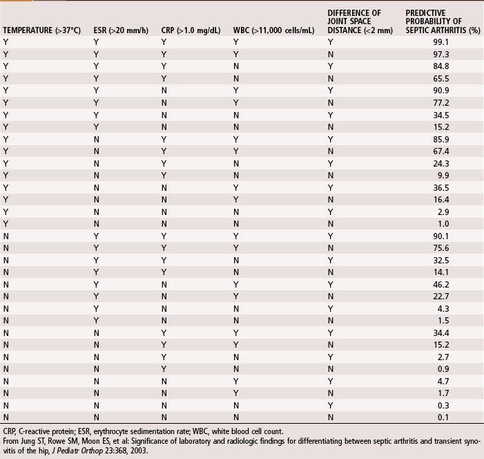
A complete blood cell count, including differential and erythrocyte sedimentation rate (ESR) and C-reactive protein (CRP), should be obtained during initial evaluation of bone and joint infections. The white blood cell count is an unreliable indicator of infection and often is normal even when infection is present. The differential shows increases in neutrophils during acute infections. The ESR becomes elevated when infection is present, but this does not occur exclusively in the presence of infection. Fractures or other underlying diseases can cause elevation of the ESR. The ESR also is unreliable in neonates, patients with sickle cell disease, patients taking corticosteroids, and patients whose symptoms have been present for less than 48 hours. Peak elevation of the ESR occurs at 3 to 5 days after infection and returns to normal approximately 3 weeks after treatment is begun. CRP, synthesized by the liver in response to infection, is a better way to follow the response of infection to treatment. CRP increases within 6 hours of infection, reaches a peak elevation 2 days after infection, and returns to normal within 1 week after adequate treatment has begun. Other tests, such as the S. aureus surface antigen or antibody test and counterimmunofluorescence studies of the urine, are promising, but their usefulness in clinical situations has not been proved. Material obtained from aspiration of joint fluid can be sent to the laboratory for a cell count and differential to distinguish acute septic arthritis from other causes of arthritis. In septic arthritis, the cell count usually is greater than 80,000/mm3, with more than 75% of the cells being neutrophils (Table 20-3). Jung et al. devised an algorithm to predict the probability of septic arthritis in children (Table 20-4). A Gram stain also should be obtained. Gram stains identify the types of organisms (gram-positive or gram-negative) in about a third of bone and joint aspirates. Intraoperative frozen section also should be obtained in cases in which infection is suspected. A white blood cell count greater than 10 per high-power field is considered indicative of infection, whereas a count less than 5 per high-power field all but excludes infection.


TABLE 20-3 Synovial Fluid Analysis


  LEUKOCYTES NEUTROPHILS (%)
Normal <200 <25
Traumatic <5,000 <25
Toxic synovitis 5,000-15,000 <25
Acute rheumatic fever 10,000-15,000 50
Juvenile rheumatoid arthritis 15,000-80,000 75
Septic arthritis >80,000 >75

From Morrissy RT: Septic arthritis. In Gustilo RB, Genninger RP, Tsukayama DT, editors: Orthopaedic infection: diagnosis and treatment, Philadelphia, 1989, WB Saunders.


Imaging Studies


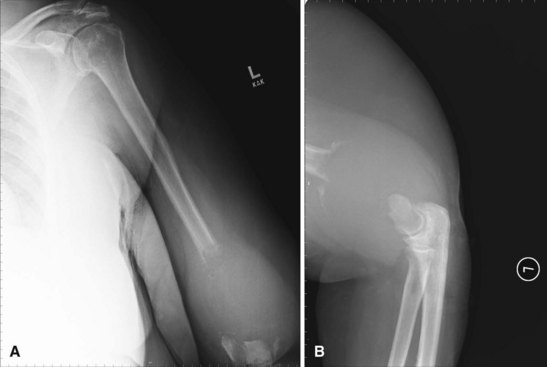
Radiographic studies are helpful but are not as useful in the diagnosis of acute bone and joint infections as they are in following responses to treatment. Plain radiographs show soft tissue swelling, joint space narrowing or widening, and bone destruction (Fig. 20-1). Bone destruction is not apparent on radiographs, however, until an infection has been present for 10 to 21 days. In addition, 30% to 50% of the bone matrix must be lost to show a lytic lesion on radiographs (Fig. 20-2). Wheat found that fewer than 5% of plain radiographs were initially abnormal in bone and joint infections, and fewer than 30% were abnormal at 1 week; however, 90% were abnormal at 3 to 4 weeks. If initial radiographs are normal in the evaluation of bone and joint infections, other imaging methods that show soft tissue swelling and loss of normal fat planes around the involved bone or joint should be used.
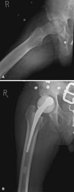


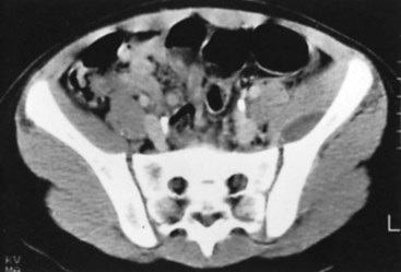
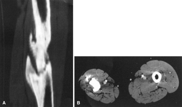
Conventional tomography can be useful in identifying a sequestrum or subchondral bony plate destruction, although it has largely been replaced with more conventional radiographic methods. Arthrography helps document proper aspiration of a suspected septic joint. Dye should be injected only after fluid is obtained from the joint because the bactericidal effect of iodinated contrast material can cause a false-negative culture result. CT can help determine the extent of medullary involvement. Pus within the medullary cavity replaces the marrow fat, causing an increased density on the CT scan. Adjacent soft tissue abscesses also are seen easily (Fig. 20-3). CT diagnosis of acute osteomyelitis is based on detection of intraosseous gas, osteolysis, soft tissue masses, abscesses, or foreign bodies. Additionally, increased vascularity after administration of a contrast agent also can aid in the diagnosis. Narrowing of the medullary cavity by granulation tissue and new bone is readily shown during the healing phase of osteomyelitis. CT identifies sequestra in chronic osteomyelitis (Fig. 20-4). It also is helpful in identifying alterations in areas poorly seen on plain films, such as the sternoclavicular joint, sacroiliac joint, and spine. Contrast material can be used to delineate abscesses in necrotic tissue that does not enhance from surrounding hyperemic tissue.


Ultrasonography can also be used to localize an abscess cavity, detect joint effusion, or guide a physician in the proper placement of the needle when obtaining aspirate from a bone or joint.


Radionuclide scanning has become a useful imaging adjunct in the diagnosis of osteomyelitis. Although radiography and CT give a structural or anatomical picture, radionuclide scanning gives a more physiological picture. Bone scintigraphy does not detect the presence of infection but, instead, reflects inflammatory changes or the reaction of bone to the infection. Radionuclide scanning also is useful in patients with metallic implants in whom CT and MRI are of limited value because of contraindications and metallic-generated artifact, although metal subtracting software is improving imaging in these patients. The three most commonly used radioisotopes are technetium-99m (99mTc) phosphate, gallium-67 (67Ga) citrate, and indium-111 (111In)–labeled leukocytes. The most common is 99mTc phosphate, which can detect osteomyelitis within 48 hours after clinical onset of infection. The uptake of this compound is related primarily to osteoblastic activity, although regional blood flow also plays a role in skeletal uptake. After intravenous injection, the technetium is distributed rapidly throughout the extracellular compartment. Bone uptake is rapid, with more than 50% of the administered dose being delivered to bone within 1 hour. The remainder of the dye is excreted by the kidneys into the urine.


The standard technique of 99mTc phosphate imaging is to perform a three-phase study. Although this does not increase the sensitivity of the test significantly, it does increase specificity from 74% to 94%. The three-phase bone scan consists of images taken in (1) the flow phase, (2) the immediate or equilibrium phase, and (3) the delayed phase. The flow-phase image is similar to a radionuclide angiogram in that it shows blood flow. The equilibrium or blood pool image shows relative vascular flow and distribution of the radioisotope into the extracellular space. The delayed-phase image generally is obtained 2 to 4 hours after injection when renal excretion has eliminated most of the isotope except that taken up by osteoblastic activity. This image shows osteoblastic activity and is positive in numerous disease states, including osteomyelitis, tumors, degenerative joint disease, trauma, and postsurgical changes. Usually a focus of osteomyelitis appears as an area of increased tracer uptake on delayed images. To have a “hot spot” on a bone scan, the vasculature to the involved bone must be intact. If blood flow to the involved area is decreased by subperiosteal pus, necrosis (i.e., sequestrum), joint effusion, vasospasm, or soft tissue swelling, a “cold” scan may result.


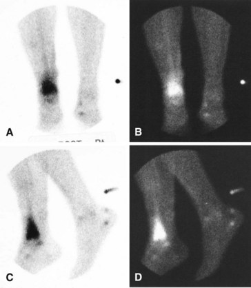
A major disadvantage of three-phase 99mTc phosphate bone scintigraphy is that the increased uptake caused by osteomyelitis is difficult to distinguish from that caused by degenerative joint disease or posttraumatic or postsurgical changes. The relative activity in each of the three phases may be helpful in differentiating other causes of increased uptake. Cellulitis causes increased activity during the flow and equilibrium phases and a decreased or normal uptake in the delayed phase. Osteomyelitis causes increased uptake in all three phases (Fig. 20-5). Increased uptake in the delayed phase but not in the flow or equilibrium phase suggests degenerative joint disease (Table 20-5). 99mTc phosphate bone scans are unreliable in neonates (<6 weeks old) and usually are negative in 60% of these patients with bone or joint infections.


67Ga citrate is the oldest tracer and has been used to localize inflammatory lesions as well as malignant tissue. The mechanism of gallium deposition is controversial; it seems to be related to increased endothelial permeability or diffusion by transportation as gallium-transferrin. The specificity of a 67Ga citrate scan alone is poor (82%). 67Ga citrate scanning can be useful in osteomyelitis when it is used in conjunction with 99mTc phosphate scanning. In purely reactive bone formation (posttraumatic or postsurgical), the intensity on the 99mTc phosphate scan is proportionally greater than that on the 67Ga citrate scan. In areas of inflammation, however, gallium uptake either exceeds that of technetium in relative magnitude or displays a different spatial configuration of activity. A disadvantage of 67Ga citrate imaging is its slow clearance after injection, which requires a delay in imaging ranging from 24 hours after injection for the appendicular skeleton to 72 hours for the axial skeleton. Specificity decreases in 67Ga citrate scintigraphy when the lesion is located peripherally rather than centrally. With the combination of 67Ga citrate and 99mTc phosphate scans, sensitivity and specificity are 70% and up to 93%, respectively, for the detection of osteomyelitis.


111In–labeled leukocytes have been suggested for differentiating between osteomyelitis and reactive bone formation. This scan is positive at earlier stages of osteomyelitis than 99mTc phosphate scintigraphy. The leukocyte scanning technique involves in vitro radionuclide labeling and injection of autologous leukocytes, predominantly polymorphonuclear neutrophilic leukocytes, followed by imaging 24 to 48 hours later. Fifty milliliters of the patient’s venous blood is obtained, separated from the other blood elements in vitro, and labeled with 111In. The labeled leukocytes are reinjected into the patient, and scans are obtained at 24 hours. A scan is positive if focal accumulation of activity exceeds adjacent normal bone activity. Scintigraphy with 111In has been reported to be helpful in the diagnosis of acute osteomyelitis, but there is disagreement about its efficacy in chronic osteomyelitis because the latter is predominantly lymphocytic and may give a negative or “cold” scan. The 111In scan also is unreliable for differentiating between aseptic and septic loosening of a painful arthroplasty. Teller et al. did not recommend the routine use of 99mTc phosphate with 111In-labeled scans for detecting aseptic or septic loosening of arthroplasty because of the high cost associated with these tests and the low specificity and sensitivity of 78% and 64%, respectively. Prandini et al., in a meta-analysis, found that 99mTc-labeled white blood cells had a greater sensitivity (89%) and specificity (90.1%) than 111In-labeled white blood cells. Several authors have recommended prolonging the scan time until 24 hours for the 99mTc-labeled white blood cell scan to improve detection. 111In-labeled monoclonal immunoglobulin is a substitute for 111In-labeled leukocytes. It seems to be as effective as 111In-labeled leukocytes, does not require phlebotomy, and avoids the risk of radiation to white blood cells and the perceived risk of malignant transformation. According to Hakki et al., compared with 111In-labeled leukocytes and 99mTc phosphate scintigraphy, monoclonal antibody fragment (LeukoScan, Granuloscint, NeutoSpect) has better sensitivity, specificity, and diagnostic accuracy. In addition, these researchers suggested that LeukoScan is a stronger diagnostic tool in patients with a low leukocyte count (i.e., human immunodeficiency virus [HIV]–infected patients) and in patients with chronic osteomyelitis. However, meta-analysis studies show that these agents are less accurate than in vitro–labeled white blood cells in most patients, and a risk of allergic reaction (some fatal) does exist especially when repeated scans are necessary. These agents have limited availability in the United States; however, the quest for a more perfect monoclonal antibody fragment continues. The detection of chronic osteomyelitis, especially of the central skeleton, can be enhanced with a 99mTc/ciprofloxacin (infection) scan and fluorine-18 (18F)-fluorodeoxyglucose-labeled positron emission tomography (FDG-PET). FDG-PET is the most accurate test (92%) with the most positive predictive value (94%). It is extremely useful for chronic infections and infections already treated with antibiotics. However, it is the most expensive and is not readily available in all health care centers.

Stay updated, free articles. Join our Telegram channel

Jun 5, 2016 | Posted by in ORTHOPEDIC | Comments Off on General Principles of Infection

Full access? Get Clinical Tree

Get Clinical Tree app for offline access