Chapter 18 Fractures of the Distal Humerus
Distal Humeral Hemiarthroplasty
Introduction
Distal humeral hemiarthroplasty (DHH), although first described in 1947, has only recently emerged as a surgical option for a number of distal humeral disorders. This has resulted from an improved understanding of the anatomy and biomechanics of the elbow,26 an increasing availability of anatomical prostheses and the recognition that DHH may provide advantages over current treatments for complex fractures of the distal humerus or pathology that destroys large segments of the distal humeral articular joint surface.
Early series reported the use of a small number of implants2–8 in a variety of pathologies, including trauma, inflammatory arthritis and tumour management. Prostheses were either custom made, non-anatomical (capitellocondylar, Kudo) or had anatomical components (Street). All these arthroplasties provided options other than open reduction and internal fixation (ORIF), interposition arthroplasty and total elbow arthroplasty (TEA)9–11 for the treatment of complex elbow pathology.
Indications
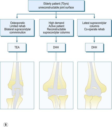
The indications for DHH are currently evolving in terms of what is appropriate and achievable, and will continue to evolve as longitudinal studies provide more clinical data. My initial experience with DHH involved younger patients with comminuted fractures of the distal humeral joint surface, sometimes combined with a column fracture, where adequate ORIF was not achievable and the attendant outcomes were unsatisfactory. In addition, with the increased use of TEA for the treatment of comminuted distal humeral fractures in the elderly patient,12–1427 it was apparent that some older patients placed too high a demand on these TEAs and DHH provided a number of advantages.
Current indications for DHH include:
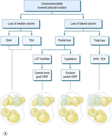
In terms of selecting whether ORIF, bone grafting, DHH, TEA or even radiocapitellar arthroplasty (RCA) is appropriate, this depends on the load-bearing columns involved, the age of the patient and the activity levels as outlined in the treatment algorithm (Fig. 18.1).


Figure 1A courtesy William PLT. Pocket picture guides to the anatomy of the joints: elbow joints. Gower Medical Publishing.
Summary Box 18.1 Indications and contraindications for DHH
| Indications | Contraindications |
|---|---|
| Acute trauma | Infection |
| Salvage of failed ORIF | Poor neurovascular status |
| Chronic malunion/non-union | Insufficient bone columns |
| Avascular necrosis of the trochlea | Loss of collateral ligaments and epicondyles |
| Tumours of the trochlea | Loss of radial head/coronoid |
Implants
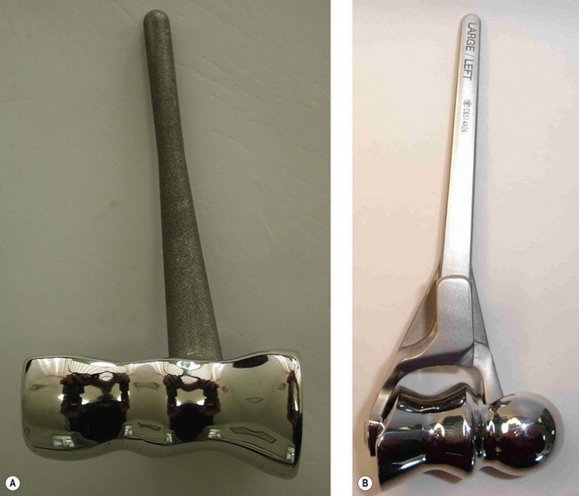
Street and Stevens6 designed a near-anatomical DHH spool in 1974. However, experience with TEA and the mechanism of humeral component loosening suggests that DHH implants should be stemmed and preferably have an anterior flange to resist joint reactive forces. There are currently two TEA systems (Fig. 18.2) available that allow a DHH (Sorbie-Questor elbow, Wright Medical Technologies; and the Latitude elbow system, Tornier) and another under development (Coonrad/Morrey DHH, Zimmer). These current prostheses have their strengths and weaknesses. Advances in the understanding of elbow anatomy and biomechanics15–18 have helped determine the correct shape and sizing of joint surfaces, along with the landmarks for appropriate implantation ensuring good stability and function.
The Sorbie-Questor19 humeral component is a monobloc component with three sizes, which allows a best fit in 95% of all elbows. Its cupped condylar shape allows for impaction up into the supracondylar columns and humeral shaft, leaving adequate space for periarticular plates for column fixation. The stem, however, is relatively short and has no anterior flange to resist anteroposterior (AP) forces; thus condylar fixation is imperative to prevent implant loosening.
The Latitude system20 has three stem sizes (with an anterior flange) and six modular anatomical spools. Although reconstruction of the epicondyles is the preferred option, a central axis hole allows suture fixation of collateral ligament repair if required. Although both systems would theoretically allow conversion to a TEA, the practicality of this is still to be demonstrated. The use of an anterior flange in humeral implant design has been shown to give better long-term humeral stem fixation and may therefore result in improved outcome for the Latitude system. At present, however, it is too early to know if this will be borne out by the long-term results.
Presentation, investigation and treatment options
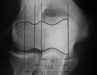
Preoperative imaging should include good AP and lateral radiographs of the elbow. In addition, since it is often unclear whether a fracture is reconstructable or will require a DHH, a CT scan should also be considered to help clarify the situation. If reconstruction is not achievable, an X-ray of the contralateral elbow will help with accurate templating (Fig. 18.3). Sizing is based on the direct correlation between the AP dimensions of the normal articular surface and the radii of the capitellum and trochlea. In addition, in the AP view, the longitudinal axis of the radial neck should align with the centre of the capitellum, while at the same time the prosthetic trochlea should align accurately with the trochlea or sigmoid fossa.


