Football


Football


Bryan T. Kelly

Ronnie P. Barnes

John W. Powell

Russell F. Warren


INTRODUCTION

Football throwers are at risk for shoulder injury secondary to both the throwing motion and the contact injury incurred by collision with another player or the ground (1, 2, 3). Although the incidence of shoulder injury in football throwers appears to be less frequent than in baseball pitchers, in our clinical experience with both professional and collegiate football athletes, we have encountered a unique spectrum of shoulder injuries including acromioclavicular (AC) and sternoclavicular (SC) joint separations, deltoid and rotator cuff contusions, shoulder dislocations and subluxations, fractures, pectoralis major injury, disorders of the biceps tendon, and overuse rotator cuff pathology (1,2,4). The risk for shoulder injury may be increased with the level of athlete (elite versus nonelite), the style of throwing (side arm versus overhead), the length of the throw, and the associated muscle fatigue that occurs throughout a game or practice. These injuries may be secondary to trauma (AC joint separation, SC joint separation, deltoid contusion, rotator cuff contusion, pectoralis major muscle injury) (1,5,6) or to chronic overuse (rotator cuff tendonitis, biceps tendonitis, impingement syndrome) (2,3).


KINEMATICS AND BIOMECHANICS

In the limited research looking at the kinematics of football throwing (7, 8, 9), there have been no reports on the muscle activation of rotator cuff muscles and shoulder synergists during the overhead football throw. Even though the football throw is similar in some respects to other overhead throwing motions, the increased weight of the football (0.42 kg versus 0.14 kg for the baseball) appears to affect shoulder position and stresses throughout the throwing motion (7,10,11). Because of the mechanical adjustments the shoulder must make to compensate for the heavier football, this throwing motion is likely to have different muscular activation patterns compared to those of other overhead throwing events. In addition, the injury patterns observed clinically are unique in this population of athletes and include traumatic injuries to the pectoralis major, AC joint, and SC joint, as well as disorders of the biceps tendon and more common rotator cuff pathology (1, 2, 3,5,6,10,11).

Phase definition and electromyographic (EMG) analysis has been thoroughly investigated for the baseball throw and relatively consistent definitions have been previously established (12, 13, 14, 15, 16). Fleisig and colleagues (17) have published one description of the phases of the football throw, but their description of the throwing motion used the same six phases that had been previously defined for baseball pitching with no regard for mechanical adjustments associated with the heavier ball. This description was made to simplify the interpretation of the results, because the purpose of the study was to compare the kinematics of the football throw and the baseball pitch (17).

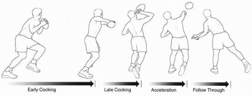
We have used video analysis of professional football quarterbacks to critically describe the phases of the football throw (18) (Fig. 22-1). Four sequential phases of the football throw were consistently observed: (a) early cocking (rear foot plant to maximal shoulder abduction and internal rotation), (b) late cocking (maximal shoulder abduction and internal rotation to maximal shoulder external rotation),(c) acceleration (maximal shoulder external rotation to ball release), and (d) follow-through (ball release to maximal cross-body horizontal adduction). The defined phases were highly consistent among the National Football League (NFL) athletes analyzed by video review, as well as among the amateur subjects tested by EMG and motion analysis (18). These phases were similar to the phases described for the baseball pitch (12,14, 15, 16,19). The average total duration of the throw was similar to what has been previously reported in the literature (1.00 ± 0.22 seconds) (9,18). Since the phases described in this study were based on discreet, functionally based extremes of shoulder motion, we were able to accurately apply these phases to all the amateur
athletes that we tested. By simply identifying the point in the throw where each of the extremes was achieved, the events that marked the transitions between phases could be reproducibly identified in all of the subjects tested. We believe that such an objective measure of phase transitions is useful to help formulate relatively consistent definitions similar to those that have been previously established for the baseball throw.


FIGURE 22-1. Four phases of the overhead football throw: early cocking, late cocking, acceleration, and follow-through. (From Kelly BT, Backus SI, Williams RJ, et al. Electromyographic analysis and phase definition of the overhead football throw. Am J Sports Med 2002;30:837-844, with permission.).

We have also looked at the EMG activity of nine shoulder muscles and correlated muscle activation throughout the defined phases of the throw (18). Our findings demonstrated that the muscle activation patterns observed were also highly consistent among athletes, and changes in muscle activation throughout the throw correlated well with the defined phases.

In his EMG analysis of the baseball pitch, Gowan and colleagues (14), defined two types of muscle activity. He defined group I muscles as those that were more active during the early and late cocking stages than during acceleration and follow-through. The muscles included in this group were supraspinatus, infraspinatus, and biceps brachii. Group II muscles were more active in the acceleration stage than in the early and late cocking stages and their activity lasted into late follow-through. These muscles included the subscapularis and the latissimus dorsi. The three deltoid muscles were minimally to moderately active throughout all four phases with no clearly discernible phases of transition. The pectoralis, although active during acceleration and follow-through, was not included in the group II musculature because the peak activities for the baseball throw was during the late cocking phase at the period of maximal external rotation.

In comparing the football throw to the baseball throw, no muscles can be defined as group I muscles, using the definitions set forth by Gowan and colleagues (14). This definition of increased muscle activation during the early and late cocking phases as compared to the acceleration and follow-through phases was not appropriate for any of the muscles tested. There were, however, based on the firing patterns, two distinct groups of muscles for the football throw. We have defined group I muscles as stabilizers (18). These muscles demonstrated relatively static levels of activity throughout the throw and included the supraspinatus, the infraspinatus, all three deltoids, and the biceps. The supraspinatus and infraspinatus were further characterized as high-level stabilizers with moderate to maximal activity throughout all four phases. The three heads of the deltoid were characterized as moderate level stabilizers with moderate activity throughout all phases. The biceps was characterized as a low-level stabilizer with minimal activity throughout all four phases. We have defined group II muscles as accelerators (18). The group II muscles for the football throw were identical to the group II muscles identified during the baseball throw: more active in the acceleration phase than in the early and late cocking phases with activity present into late follow-through. The muscles included in this group were the subscapularis, the pectoralis major, and the latissimus dorsi. These muscles provided the majority of the force that was imparted into the football throw.

The presence of persistently high levels of activation of the three accelerator muscles into the follow-through phase may not be intuitively understood. All three of these muscles are internal rotators of the humerus and hence should not be expected to decelerate the internal rotation forces of the humerus as is required during the follow-through phase. However, two considerations may help to explain this phenomenon. With regard to the subscapularis, this muscle contributes to the normal co-contraction forces of the rotator cuff that are essential for maintaining the humeral head centered on the glenoid. Although the subscapularis functions
as an accelerator during the acceleration phase, it has a dual role with the remainder of the rotator cuff as a cocontractor during the powerful joint distraction forces experienced during the follow-through phase. The latissimus dorsi and pectoralis major muscles may be recruited to provide additional co-contraction forces to reinforce the job that is routinely controlled by the rotator cuff musculature alone during less forceful activities. The high kinetic forces that are experienced across the shoulder joint during football throwing may not be adequately countered by the rotator cuff alone. Thus, during forceful overhead activities such as football throwing and baseball pitching, the latissimus dorsi and pectoralis major muscles may be required to control further distraction across the shoulder joint during the follow-through phase.

In comparing the kinematics and kinetics between the baseball pitch and the football pass, Fleisig and colleagues (7) demonstrated several differences between these two overhead activities. Most notably, during arm deceleration, pitchers produced greater forces and torques in the shoulder and elbow. They also demonstrated that higher arm speeds were generated in pitching. Shoulder internal rotation velocities were between 3 and 4.5 times faster during the baseball throw and elbow extension velocities were between 2 and 3 times faster during the baseball throw (7,9). In addition, greater degrees of shoulder abduction and external rotation were achieved during the baseball throw (B. Kelly, unpublished data). In further evaluating the baseball EMG work by Gowan and colleagues (14), it is notable that the accelerator muscle group demonstrated considerably more activity during the baseball throw compared to what was found during the football throw in this study. These findings are consistent with the theory that the accelerator muscles have a dual responsibility to provide additional cocontraction force during the follow-through phase to further stabilize the shoulder and prevent joint distraction. Since the baseball throw results in greater kinetic forces across the shoulder joint during deceleration (7), it is appropriate to see the greater levels of muscle activation in the accelerator group during baseball pitching compared to football throwing (14).

Clearly defining different types of muscle activation patterns has clinical implications. First, by knowing the manner in which different muscles fire during the throw, athletes can be given both sport- and muscle-specific conditioning protocols. The most effective training method for optimal conditioning of the stabilizer muscles should differ from the most effective conditioning of the accelerator muscles. Stabilizer muscles may benefit from more isotonic conditioning while accelerator muscles may be more effectively strengthened with plyometric and acceleration exercises. Knowledge of these two different muscle groups also provides insight into rehabilitation protocols. The goal of rehabilitation of injured muscles should be the return to sport-specific kinematics. If the mode of activation during the throw can be more accurately simulated during rehabilitation, we would anticipate a quicker return to full functional activity (20, 21, 22, 23). Additional clinical correlation and investigation is warranted to confirm these hypotheses.

Ultimately, we seek to identify which anatomic structures are most important to the football throwing motion. Just as earlier EMG studies led to the development of sports-specific preventive and therapeutic protocols, EMG analysis of the shoulder musculature during the football throw will lead to a better understanding of throwing injuries associated with football throwing (12,23, 24, 25) and more specific rehabilitation and conditioning programs will be developed that might better protect quarterbacks from the development of shoulder conditions. By further identifying associated risk factors for shoulder injury, additional safety measures and precautions can be more intelligently exercised.


ETIOLOGY OF INJURY

We have accessed the NFL Injury Surveillance System (NFLISS) to identify all injuries to quarterbacks that have been reported to the NFL between 1980 and 2001. The injury data collected in the NFLISS is based on the primary clinical impression of the clinical diagnosis made by the medical staff involved. The data reflect only those cases reported during the season (training camp to Super Bowl) and that required the player to be restricted from playing for at least 2 days. Over 22 seasons, 1,534 quarterback injuries were reported to the NFLISS with a mean of 18.8 days of playing time lost. Most of these injuries (83.8%) have occurred during a game, with slightly more occurring on grass (55.7%) compared to turf (44.3%). Passing plays are responsible for 77.4% of all quarterback-related injuries.

Of the 1,534 injuries reported, 233 (15.2%) involved the shoulder, including the glenohumeral joint, proximal humerus, scapula, clavicle, AC joint, SC joint, long head of the biceps tendon, rotator cuff, scapular stabilizers, deltoid, and pectoralis major muscles (Tables 22-1 and 22-2). Shoulder injuries were the second most common injury sustained by quarterbacks, following closely behind head injuries (15.4% of reported injuries). The most common mechanism of shoulder injury is related to direct trauma, either from contact with another player or with the ground (80.3%). Nearly 70% of shoulder injuries occurred while the quarterback was being tackled: 47.3% occurred while being tackled as the passer, 12.6% while being tackled as the ball carrier, and 9.6% while being tackled after the pass. Only 12.4% of injuries were reported as being secondary to the actual throwing motion.

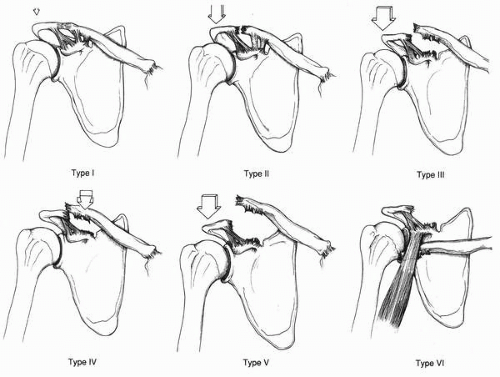
Overall, the most common single shoulder injury identified was an AC joint sprain incurred while being tackled or during a collision with another player or the ground (39.6%). Of all AC joint sprains, 44% were type I, 24%
were type II, 20% were type III, and 12% were not specified. The second most common group of injuries was shoulder contusions from collisions or tackling and involved the deltoid (10.9%), rotator cuff (8.4%), and scapular stabilizers (1.7%). Other injuries reported as a result of direct trauma included anterior shoulder dislocations (8%), fractures of the proximal humerus, scapula, and clavicle (3.7%), posterior shoulder dislocations (2.5%), SC joint sprains (2.5%), impingement and bursitis (1.7%), pectoralis major injuries (1.2%), biceps tendonitis (0.4%), and axillary nerve injury (0.4%).


TABLE 22-1. INJURIES TO THE SHOULDER AND CHEST IN PROFESSIONAL QUARTERBACKS REPORTED TO THE NFLISS FROM 1980 TO PRESENT


Injuries to Shoulder and Chest


Overall


Tackled/Collision


Throwing


Other


Deltoid contusion


11.30%


10.90%


0%


0.40%


Rotator cuff contusion, sprain, tendonitis


18.10%


8.40%


6.10%


3.60%


Scapular, trapezius contusion


1.70%


1.70%


0%


0%


Anterior shoulder dislocation, subluxation, capsulolabral injury


8.40%


8.00%


0.40%


0.00%


Posterior shoulder dislocation, subluxation, capsulolabral injury


2.90%


2.50%


0.40%


0%


Synovitis, capsulitis


1.60%


0.00%


0.80%


0.80%


Nerve injury


0.40%


0.40%


0%


0%


Biceps tendinitis


4.30%


0.40%


3.50%


0.40%


Impingement, bursitis


2.90%


1.70%


0.40%


0.80%


Fracture (humerus or scapula)


1.20%


1.20%


0%


0.00%


Pectoralis major tear, sprain


1.20%


1.20%


0%


0%


54.00%


36.40%


12%


6%


The most common injury identified as a result of the throwing motion itself was rotator cuff tendonitis (6.1%) followed by biceps tendonitis (3.5%). Other injuries reported as being associated with the throwing motion itself included synovitis and capsulitis (0.8%), anterior labral tear (0.4%), posterior capsular strain (0.4%), impingement (0.4%), and SC joint strain (0.4%). The clinical spectrum of injuries observed in the NFLISS are consistent with the kinematic and EMG findings reported previously (18). Compared to the baseball throw, the football throw is associated with significantly slower rotational velocities, decreased extremes of range of motion, and decreased electrical activity from the rotator cuff and surrounding shoulder musculature (7,9,14,18). Based on these biomechanical data, one would expect fewer problems with chronic overuse types of injury such as those experienced much more commonly in baseball pitchers. This is, in fact, what is observed, with most shoulder injuries occurring as a result of trauma (80%) and less than 15% resulting from the actual throwing motion.


TABLE 22-2. INJURIES TO THE CLAVICLE, AC JOINT, AND SC JOINT IN PROFESSIONAL QUARTERBACKS REPORTED TO THE NFLISS FROM 1980 TO THE PRESENT


Injuries to Clavicle/AC joint/SC joint


Overall


Tackled/Collision


Throwing


Other


AC Joint Sprain


40.00%


39.60%


0%


0.40%


SC Joint Sprain


3.40%


3%


0.40%


0%


Clavicle Fracture


2.50%


2.50%


0%


0%


45.90%


45.10%


0.40%


0.40%


OVERVIEW OF SPECIFIC INJURIES


Acromioclavicular Joint Separation

Acute injuries to the AC joint are common in a variety of contact sports and are the most common isolated injury to the shoulder complex in quarterbacks (40% of reported shoulder injuries in the NFL). The typical mechanism for an acute traumatic injury is a fall onto the shoulder or a direct blow to the acromion while being tackled during or after passing. A shoulder separation occurs when there is sufficient energy to cause a partial or complete disruption of the AC or coracoclavicular (CC) ligaments. Acromioclavicular joint dislocations have been classified into six types based on the extent of ligament damage (26, 27, 28, 29, 30) (Fig. 22-2). Type I injuries involve a sprain of the AC ligament with intact AC and CC ligaments. Type II injuries are the result of more severe trauma, resulting in disruption of the AC ligaments but leaving the CC ligaments intact. Type III
injuries involve disruption of both the AC and CC ligaments, which results in visible dislocation of the AC joint with displacement of 50% to 100%. Although there may be mild elevation of the distal clavicle, the more significant cause of the deformity is from depression of the dissociated shoulder girdle. Types IV, V, and VI injuries are the result of more violent forces leading to complete disruption of the surrounding ligaments and musculature. Type IV injuries involve posterior displacement and penetration of the trapezius muscle by the distal clavicle. Type V injuries are characterized by marked superior displacement with greater than 100% increase in CC distance. In type VI injuries, the distal clavicle dislocates to a subcoracoid position (28).


FIGURE 22-2. Classification of acromioclavicular joint separations. (From Rockwood CA, Williams GR, Young DC. Disorders of the acromioclavicular joint. In: Rockwood CA, Matsen FA, eds. The shoulder, 2nd ed. Philadelphia: WB Saunders, 1998:483-553, with permission.).

Patients with type I and II AC dislocations have pain and swelling over the superior shoulder, focal tenderness at the AC joint, and pain with cross-arm adduction. Type I injuries have no displacement of the clavicle, whereas in type II injuries, prominence of the joint is present with a radiographic displacement of up to 50%. Type I and II AC separations generally require only symptomatic care. Strengthening programs, including range-of-motion exercises, can usually be initiated within 1 to 2 weeks after the acute pain has subsided. Athletic participation can usually be resumed within 3 to 4 weeks, once normal strength and range of motion has returned (27). Nonsurgical treatment of types I and II injuries leads to good results in more than 90% of cases, although posttraumatic degenerative joint disease may sometimes develop secondary to meniscus or articular cartilage damage at the time of injury. If pain persists, it is reasonable to perform a Mumford procedure with expectation of a good result and ultimate return to full preinjury level of activity (31).

The management of type III injuries remains somewhat controversial (11). In 1974, Powers and Bach (32) reported that 92% of 116 type III injuries were managed operatively. Seventeen years later, Cox (33) reported that most surgeons (72% of residency chairmen and 86% of team physicians) were managing type III injuries nonoperatively (34). Currently, there is much support in the literature for the nonoperative treatment of type III injuries in nearly all patients (34, 35, 36, 37). However, disagreement remains regarding the appropriate management of the elite throwing athlete (11,35,37,38). McFarland and co-workers (35) surveyed 42 orthopedic surgeons representing 28 major league baseball teams concerning treatment of type III AC separations in professional throwing athletes. In this survey, 31% of the surgeons recommended immediate operative intervention and 69% believed that nonoperative management would be
more appropriate. Twenty-five of the orthopedists surveyed had actually treated type III AC separations in throwing athletes and reported that 80% of the patients treated nonoperatively regained normal function and achieved complete relief of pain, and 90% had normal range of motion after treatment. Of those treated operatively, 92% regained normal function, achieved complete relief of pain, and had normal range of motion after surgery. Absolute treatment guidelines for the elite throwing athlete cannot be made based on the available literature in that operative and nonoperative treatment appear to give similar results (35), although some authors maintain that type III AC joint sprains may result in an alteration in throwing mechanics that can manifest as subacromial impingement and should therefore be addressed surgically (34).

As has been previously explained, the football throwing athlete must be considered differently than the baseball throwing athlete because their associated injuries are more commonly caused by traumatic force than by overuse. Cardone and colleagues (39) reviewed 14 Australian Rules Football (ARF) players who were seen consecutively by a single surgeon with grade III AC joint injuries. Treatment of this group may be more analogous to the situation encountered with quarterbacks. In this group, eight players elected for nonoperative management and six for operative management. Two players in the nonoperative group subsequently underwent surgical reconstruction after failure of nonoperative treatment. The mean return time to noncontact training was 2.4 weeks (range 1 to 4, SD 1.52) in the nonoperative group and 6.3 weeks (range 3.5 to 10, SD 2.99) in the operative group. However, return to sports-specific training (contact training) was at a mean of 20.8 weeks (range 10 to 32, SD 8.56) in the nonoperative group and 13.6 weeks (range 6 to 24, SD 7.06) in the operative group. Although limited by the small numbers, the results show a trend toward faster return to ARF and a more satisfactory outcome for patients undergoing surgery compared to their nonoperative cohorts.

Initial surgical intervention is usually indicated for the treatment of type IV, V, and VI injuries because of the severe displacement of the clavicle. Popular operative techniques described for reduction of the AC joint include fixation across the AC joint, dynamic muscle transfer, and fixation between the clavicle and the coracoid (34). Reconstruction of the ligament can be performed using the modified Weaver-Dunn procedure, which involves limited distal clavicle resection, transfer of the coracoacromial ligament, and stabilization of the coracoclavicular interval with suture, soft tissue, or hardware. The postoperative regimen often requires up to 6 weeks in a sling, followed by motion and strengthening exercises (27). The modified Weaver-Dunn has had favorable results in reducing pain and improving function in athletes with symptomatic type III, IV, and V acromioclavicular dislocations (40).

Verhaven and co-workers (41) prospectively followed 18 consecutive athletes with an acute type V AC sprain treated with a coracoclavicular repair using a double velour Dacron graft. All patients were reviewed after a mean follow-up period of 6 years (range 2 to 9 years). At follow-up, 12 patients (66.7%) showed a good or excellent result according to the Imatani evaluation system, and six patients (33.3%) demonstrated a fair or poor result according to the same system. Loss of reduction was encountered in eight shoulders (44.4%) despite an initial anatomic reduction. No correlation was seen between the overall scores at follow-up and the degree of residual dislocation, between the overall scores and the presence of coracoclavicular calcifications or ossifications, between the overall scores and the development of posttraumatic arthritic changes, or between the overall scores and the presence of osteolysis of the distal clavicle. These authors concluded that surgical treatment for acute type V AC separations in athletes provides fewer good results than similar treatment for type III lesions. However, in this series, all patients returned to the same level of sport activity as before injury (41).


Authors’ Preferred Approach

Grade I and II injuries, as noted, are treated nonoperatively with a progressive rehabilitation program. Similarly, after initial protection with a sling, most grade III injuries in athletes are placed in a rehabilitation program and observed. Surgery has rarely been necessary: there is little in the literature to support early repair even in a quarterback. Taft (42) previously reported that about 10% of nonoperative AC joint injuries go on to need surgical care. In addition, most strength studies have failed to show significant deficits with nonoperative treatment in throwers. We have treated several NFL quarterbacks nonoperatively with a grade III separation who returned to play at 5 or 6 weeks.

In contrast, grade IV, V, and VI injuries need surgical treatment. With gross displacement as in a grade V injury, the standard Weaver-Dunn procedures appear to be inadequate in some patients. It appears that the instability is not only inferior but that the acromion migrates medially, which places excess stress on a simple coracoclavicular graft. Thus, we have preferred to use an autograft or allograft to reconstruct both the coracoclavicular ligament and the acromioclavicular ligaments (Fig. 22-3). In addition, when the joint has been fractured, we prefer to excise the distal clavicle and repair the ligaments and muscular attachments. This may be sufficient in acute cases but if the tissue is poor or the injury is old, then a reconstruction of the acromioclavicular as well as the coracoclavicular ligaments is indicated. In a limited number of these cases, we have noted improved maintenance of stability.


Rehabilitation Pearls

Acute AC sprains involving all classifications can be painful. Early immobilization is prescribed for most type III
injuries. Acute nonsurgical management of these conditions includes pain management, reduction of swelling, and maintaining shoulder function and overall range of motion. Pain and swelling are the primary inhibitors to increased motion and initiation of progressive resistive exercises. Frequent applications of ice packs are helpful in pain control. Strengthening programs should include specific exercises for the deltoid muscles. Forward elevation to beyond 90 degrees or more is encouraged. Throwers with AC injuries to the shoulder progress in rehabilitation from Codman’s and pendulum activities to more aggressive range-of-motion exercises as pain and swelling permits. Focus on strength, endurance, proprioception, and neural control can be achieved only as pain and swelling decreases. Functional activities involving throwing are performed only in the final phases of rehabilitation. Return to throwing in sports must be based on pain-free range of motion and healing of the joint with normal mechanics. Athletes who return to play with some lingering point tenderness may need a bridged hard shell protective pad to protect the area from direct impact while playing.


FIGURE 22-3. Autograft or allograft reconstruction of the coracoclavicular and acromioclavicular ligaments.


Sternoclavicular Joint Separation

Injury to the SC joint in quarterbacks is much less common than AC joint injuries, accounting for less than 3% of all reported shoulder girdle injuries in the NFLISS. The decreased incidence is related to the medial location of the joint, although indirect forces can be transmitted through the shoulder girdle, resulting in sprains, fractures, or dislocations (43). Difficulty in diagnosis is related to the relative rarity of the injury as well as limitations of standard radiographic views. Radiographic examination should include standard anteroposterior (AP) and lateral chest radiograph looking for asymmetry of the SC joint compared to the contralateral side (43). Rockwood (44) described the “serendipity view” obtained with a 40-degree cephalic tilt, which reveals a true caudocephalic view of both SC joints and the medial clavicles. In an anterior dislocation, the affected clavicle appears superior and in a posterior dislocation it appears inferior (44). Acute sprains of the SC joint are classified as type I, type II, or type III, depending on the degree of injury to the supporting ligaments and joint capsule (43). In a type I injury, there is a partial disruption of the ligaments but the clavicle remains in its anatomic position with respect to its medial articulation. In type II injuries, there is an incomplete disruption of the ligaments and some degree of subluxation can occur in either the anterior posterior direction. Symptomatic treatment, consisting of local modalities, analgesics, and short-term immobilization, is preferred for type I and II injuries (45).

A type III sprain or dislocation results from a higher energy injury and produces a complete disruption of the supporting ligamentous structures. Most traumatic SC dislocations are anterior in direction, resulting from a posteriorly directed force to the anterolateral aspect of the shoulder. Clinically, localized pain and prominence over the medial end of the clavicle are present, and CT scans are useful in further evaluating the direction of the injury and the proximity of the injury to surrounding neurovascular structures (27). Closed reductions may be attempted, but are often difficult to maintain with immediate redisplacement of the medial clavicle, and sling immobilization is usually required for a minimum of 2 to 3 weeks (27,43). Nonsurgical management results in good functional outcomes in most cases (46), and open reduction of anterior dislocations is rarely indicated (43).

Because of the strong forces involved and the proximity of the joint to the great vessels and other mediastinal structures, posterior sternoclavicular injuries can be serious and potentially life threatening (47, 48, 49). The injury is typically a result of a direct blow to the anteromedial aspect of the clavicle or of an anteriorly directed force to the posterolateral aspect of the shoulder. Signs of dyspnea, dysphagia, venous congestion, or hemodynamic instability indicate possible compression or compromise of nearby mediastinal structures and require emergent reduction (47,48). Marker (50) reviewed the reported cases of posterior dislocation of the sternoclavicular joint and found only 100 cases since 1824. In reviewing the published reports, he found that this injury is seen particularly in connection with American football and that there is often a delay in diagnosis with potentially serious complications. Closed reduction of these injuries should be performed within 7 days by abduction and extension of the shoulder and manual manipulation of the clavicle. Rockwood (44) described a technique of reduction under general anesthesia in which the clavicle is percutaneously grasped with a towel clip to facilitate reduction. If
closed reduction fails, the reduction is not stable, or the dislocation is greater than 7 days old, open reduction and stabilization is indicated (43).

Only gold members can continue reading. Log In or Register to continue

Stay updated, free articles. Join our Telegram channel

Sep 16, 2016 | Posted by in ORTHOPEDIC | Comments Off on Football

Full access? Get Clinical Tree

Get Clinical Tree app for offline access