Chapter 25 Foot orthoses
The benefit of a foot orthosis is subject to much discussion and controversy.7 Instrumentation has been used to conduct studies measuring its direct effects on the foot. Quantifying its indirect effects on proximal joints has proven to be difficult, so little sound research is available. As technology improves, research likely will show the functional benefits of foot orthoses providing the necessary documentation for insurance coverage. In the United States, the Medicare health care system presently funds foot orthoses only when they are used for treatment of patients with diabetes. Even without clear documentation on the effectiveness of foot orthoses, these devices are commonly prescribed for treatment of various foot/ankle pathologies, and many individuals profess their benefits.
Evaluation
Skin condition provides some insight into the cause of the problem and assists with orthotic design selection. Callus formation is a result of repetitive pressure. The location of the callus on the plantar surface pinpoints areas of high stress when weight bearing. This information is used when designing the orthosis to dissipate the stress in these areas. Callus forms not only on the plantar surface but on any stressed area. The lack of width and depth of the shoe’s toe box may result in excessive pressure over the dorsum of the foot. In some cases, callus formation is the source of discomfort and patient education will be important in controlling callus buildup. Dry cracking skin may be the result of a systemic condition. Delicate fragile skin will require softer materials in the construction of the orthosis. Corrective forces applied by the orthosis must be limited to protect skin integrity. Sensation is an important factor in protecting the overall integrity of the foot, and any deficit in its normal function must be noted. Diminished or complete absence of sensation will require a more accommodative and protective approach by using an orthosis made of softer materials. Careful examination of the skin is essential in preventing complications and providing optimum orthotic treatment.
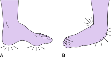
Shape of the foot (cavus or planus) is a good predictor of potential problems that commonly present (Fig. 25-1).3 Cavus feet usually are less flexible, resulting in decreased shock-absorbing capabilities. This deficit in the mechanics of the foot results in excessive pressure on the ball of the foot and the heel. Severe cavus deformities also result in pressure on the base and the head of the fifth metatarsal. At initial contact and loading response, the cushioning effect that occurs through pronation when the talonavicular joint and the calcaneocuboid joint axis are congruent is lost. The foot remains stiff throughout stance phase, with normal performance through the latter part of stance when a rigid toe lever is necessary for normal push-off.10 In contrast, the planus foot usually is flexible and presents with problems related more to poor alignment of the joints of the foot and ankle. With the foot in the pronated position the calcaneus remains everted, the talus is plantarflexed, and the forefoot is abducted.8 Stress is placed on the supporting structures of the medial arch, and severe pronation may result in stress being placed on the lateral malleolus. Through the stance phase the foot acts relatively normal during initial contact and loading response as the foot absorbs the shock, but additional effort is necessary to achieve the rigid toe lever necessary for the latter part of stance phase. Although there is a potential for the mentioned deformities to develop, many individuals are asymptomatic and never require professional treatment. The general shape of the foot has an effect on how the foot responds to different supportive approaches.
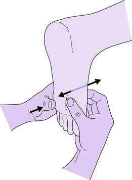
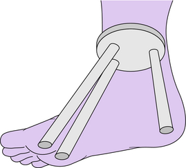
To completely assess foot function, ROM in the foot and the ankle must be determined. The hindfoot, midfoot, and forefoot are assessed in both the open and closed chain environment to determine if any limitations or excesses are present. Emphasis is placed on the closed chain, when the role of the foot is most important. Normal subtalar joint ROM permits the foot to operate naturally moving from pronation at the beginning of stance to supination during the latter part of stance phase.10 If limitations exist, the foot will not perform normally throughout the stance phase. Several techniques are available to determine subtalar neutral. Whatever the method used, the clinician should develop consistency in finding this position. With the foot held in a subtalar neutral position,15 the fifth metatarsal is dorsiflexed to lock the midfoot joint (Fig. 25-2). In this position, note the alignment of the calcaneus to the long axis of the tibia, documenting any significant abnormalities. Also note the flexibility and position of the first ray. Some research findings point to the importance of first ray stability.12 If the foot is viewed as a three-legged stool, a flexible first ray represents an instability in the stool (Fig. 25-3). Following this same concept, the foot will have a greater tendency to pronate and/or more stress will be placed on the second metatarsal to stabilize the foot. If the first ray is stable, its alignment with all the metatarsals should be noted to determine if some compensation is required to distribute the weight evenly across the heads. True ankle ROM must be determined to ensure that the foot can perform normally during gait (see Chapter 23 for a review of ROM examination and anatomy). In normal gait the ankle dorsiflexes 5 to 10 degrees. Significant limitations to this range will change the normal biomechanics of the foot, resulting in some compensatory changes (Fig. 25-4). Under these circumstances more pressure is placed on the metatarsal heads during the late part of midstance, with additional stress placed on the midfoot. Inspection of the metatarsophalangeal (MTP), proximal interphalangeal (PIP), and distal interphalangeal (DIP) joint ROM of the toes will reveal existing complications as well as the development of hammer toe or claw toe deformities.
Manual muscle testing of the major muscle groups surrounding the foot and ankle provides important information regarding weaknesses that may be the source of foot complications. The ankle dorsiflexors work as an antagonist to the plantarflexors, assist with toe clearance through swing, ensure heel contact at the beginning of stance phase, and prevent foot slap at loading response.10 Weakness of this group usually requires an ankle–foot orthosis to compensate for the lost function. Roles of the plantarflexor muscle group include antagonist to the dorsiflexors, weight acceptance during midstance, and stability of the toe lever during terminal stance.14,16 Absence of this muscle group warrants use of an ankle–foot orthosis with a solid ankle or dorsiflexion stop. Inversion and eversion strength play an important role in hindfoot and midfoot function. Weakness in the function of the inverters and everters will result in mediolateral instability. One significant role of these groups is to ensure good alignment and stability in preparation for initial contact and loading response. To resist excessive varus or valgus tendencies during loading response, heel wedges and posts can be added to the foot orthosis.5,9,13 Attention should focus on the function of the posterior tibialis given its importance in medial longitudinal arch stability and plantarflexion.2 During the muscle testing process, discomfort could be a sign of injury to the muscle, attachment points, or tendons. Muscle imbalances at the MP, PIP, and DIP joints result in the development of toe deformities and should be noted. To ensure successful orthotic treatment, manual muscle testing of the foot and ankle should be performed to determine the most appropriate orthosis to be prescribed.
The most important role of the foot is weight bearing during ambulation. Therefore, the evaluation should include observation of foot function during weight bearing and walking. Some important observations related to foot function include the amount of time spent on each extremity, ankle motion, calcaneal motion, pronation and supination, and tibial internal and external rotation. Findings should be compared to normal gait10 and any abnormalities documented. Identifying the phase of gait at which the pain or problem is present allows the clinician to isolate the problem and to examine any deficits present during that stage.


