Chapter 126 Extensile Surgical Exposures for Revision Total Knee Replacement
Adequate exposure is one of the most common difficulties encountered in revision knee arthroplasty. Choosing the correct surgical approach while having a comprehensive understanding of the anatomic pitfalls allows the surgeon to proceed safely and achieve a successful outcome for the patient.36 Wound edge necrosis and extensor mechanism rupture are significant complications that can adversely affect the outcome of revision knee surgery.29 Such complications can be avoided with judicious preoperative planning and knowledge of safe approaches to exposing a failed knee arthroplasty. Great care must also be taken in protecting the collateral ligaments, which are at particular risk at the level of the joint line and posterolaterally, where neurovascular structures are in close proximity to the joint. Careful attention must be paid to those patients with previous incisions, because they are at risk of wound breakdown; it can also be particularly difficult to gain adequate exposure in those with restricted range of movement. Other comorbidities may have implications as well, such as vasculitis, which can occur with polyarthropathies such as rheumatoid arthritis. These patients are at risk of poor wound healing and skin edge necrosis. The risk of deep infection is further increased in patients with renal failure, acquired immunodeficiency syndrome with a CD4 count of 200 or lower, diabetes, psoriasis, and rheumatologic conditions such as rheumatoid arthritis and systemic lupus erythematosus.42
Anatomy
The blood supply to the skin overlying the knee is well understood because of the results of a cadaveric study conducted by Haertsch and associates.16 Anastomosis of blood vessels immediately superficial to the deep fascia that are fed by perforating vessels from below the fascia is noted. From this anastomosis, blood vessels course their way through the subcutaneous fat and supply the dermis and epidermis. No such anastomoses are seen within the dermal layers; hence, extensive dissection superficial to the deep fascia will disrupt the blood supply to the skin, increasing the risk for areas of ischemia and necrosis. Therefore, if most of the dissection in exposing the knee is kept below the fascial layer, the skin’s blood supply is less likely to be compromised.
The origin of the blood supply to the skin overlying the knee is asymmetrical. Perforators arise mostly from the medial aspect of the knee from the saphenous artery and the descending geniculate artery. If a midline incision is already present, this should be used for the revision. Although this is the situation in most cases, other incisions may occasionally be present. A previous transverse skin incision obviously cannot be avoided and should be crossed at 90 degrees.40 However, creating a skin flap with an acute angle of less than 60 degrees at the intersection of the incisions risks compromising the blood supply and should be avoided. If a previous oblique incision is present, it should be crossed at as close to 90 degrees as possible, and the incision can be curved away from the intersection if necessary.
If multiple longitudinal incisions are in close proximity, then the most lateral incision should be used, while preserving the blood supply medially.7 If this is not possible, then the surgeon should aim to leave an intact skin bridge of approximately 6 cm between the previous incisions and the new one.
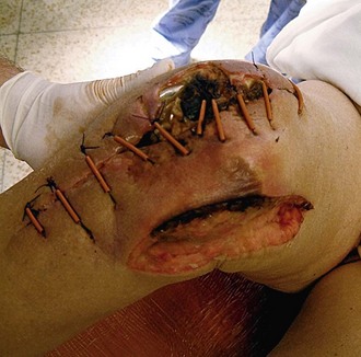
The area of skin that is particularly vulnerable during revision surgery is the anteromedial aspect of the proximal tibia, because of limited soft tissue cover deep to the skin. Careful attention should be paid to this area postoperatively and rapid intervention instigated should problems arise because of the risk of deep infection (Fig. 126-1). Should skin necrosis occur, skin grafting alone is rarely successful because of the limited muscle coverage. In this scenario, a medial gastrocnemius rotation flap is a good option when additional soft tissue coverage is required.* This should be done as early as possible, because delay may lead to infection.
The blood supply to the patella forms a plexus of arteries surrounding it supplied by the descending geniculate, superior medial, inferior medial, superior lateral, and inferior lateral geniculate arteries and the anterior tibial recurrent artery.18,19,34 From this plexus, branches arise in front of the patella; these include the infrapatellar artery and the oblique prepatellar artery, which is found within the infrapatellar fat pad. The distal pole of the patella receives its blood supply inferiorly,34 and the remaining patella receives its blood supply from vessels that penetrate it over the middle third of its anterior surface. The standard medial parapatellar approach will interrupt the contribution from the medial vessels to the plexus, and excision of the lateral meniscus at the index operation will compromise the inferior lateral geniculate artery. Similarly, when the infrapatellar fat pad is excised, the superior lateral geniculate artery18 and the branches of the recurrent anterior tibial artery may be interrupted. If a lateral release has also been performed, then the blood supply to the patella can potentially become somewhat tenuous. An increased incidence of patellar avascular necrosis with its subsequent fragmentation has been observed with lateral release and sacrifice of the superior lateral geniculate artery.32 The superior lateral geniculate artery is found running horizontally and immediately distal to the inferior border of the vastus lateralis muscle. When a lateral release is performed from within the knee joint, great care must be taken to try to preserve this vessel and thus avoid compromising the blood supply to the patella. It is advisable to avoid a release from “outside-in” because this involves considerable undermining of the lateral skin edge with the inherent risk of causing skin edge necrosis.
Identifying the collateral ligaments, as well as the capsular structures found laterally, medially, and posteriorly, is an important step in the surgical exposure of revision knee surgery.18 The medial collateral ligament is a flat, triangular band that courses its way from the medial femoral epicondyle, just distal to the adductor tubercle, and inserts into the tibia 2 cm distal to the joint line. Its anterior margin forms the vertical base of the triangle; the posterior apex of the triangle blends with the capsule and is attached to the medial meniscus. Above its distal attachment, the ligament is crossed by the tendons of sartorius, gracilis, and semitendinosus with a bursa interposed. The superficial medial collateral ligament, along with the pes anserinus, inserts more distally. The semimembranosus tendon inserts into the posterior aspect of the medial tibial condyle. From this attachment, it gives off three expansions. One passes anteriorly along the medial surface of the condyle deep to the deep medial collateral ligament. The second expansion passes obliquely and superiorly to the lateral femoral epicondyle and is called the oblique popliteal ligament. The third expansion forms a thick, strong fascial layer overlying the popliteus and inserts into posterior aspect of the tibia along the soleal line.
Knee flexion offers no protection for the popliteal artery, as it remains tethered to the posterior capsule at the level of the knee joint.43 From the neurologic point of view, the peroneal nerve is at greater risk during revision surgery than the tibial nerve. Damage to the peroneal nerve can occur from traction (especially if the normal mechanical axis is restored in a valgus knee), compression, and laceration. The common peroneal nerve descends on the lateral aspect of the joint and initially is medial to the biceps tendon; it then courses just behind its insertion on the fibular head.36 It is at particular risk from lateral release or during release of the biceps tendon; for this reason, such a procedure is best avoided.
Preoperative Assessment
History
The history should include a review of any possible wound healing problems, previous nerve injury, or weakness of knee extension, which may suggest disruption of the extensor mechanism. A history of knee stiffness should precipitate further questioning as to the duration of stiffness or loss of motion, as this can affect the choice of surgical approach used. Specific questions regarding infection should be asked, specifically if the knee wound from the primary operation took a long time to heal, or if it was complicated by prolonged drainage or a “superficial” infection. Within the systemic inquiry, information regarding peripheral vascular disease is helpful, as it may point to other possible causes of the patient’s pain, but it is also relevant as to the risk of developing wound necrosis and a postoperative ischemic limb.28
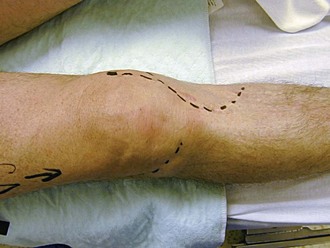
During the examination, the location and shape of surgical scars should be carefully assessed (Fig. 126-2). The general health of the skin and its capillary return in the vicinity of the planned incision should be reviewed. Discoloration of the wound edge at the previous incision with hemosiderin may suggest previous wound healing problems. The range of motion should be carefully checked before any revision knee replacement is performed. A stiff knee will likely require extensile maneuvers to achieve safe exposure. Any patient with less than 70 degrees of flexion is a likely candidate for extensile exposure.

Figure 126-2 Preoperatively, all surgical incisions should be clearly marked with an indelible marker.
A knee that has been infected is associated with increased scar formation resulting in stiffness, which can also increase the risk of patellar tendon avulsion.15 Neurovascular status should be inspected, and if there is any doubt as to the blood supply distally, additional imaging studies should be requested, along with the opinion of a vascular surgeon. Poor venous return can also be a problem and may cause tissue ischemia and wound breakdown secondary to venous engorgement. If any wound issues are envisaged, preoperative review by a plastic surgeon can be timely. Some plastic surgeons may advise creating a flap before the revision is undertaken, although this is uncommon. Occasionally, tissue expanders can be used to increase the amount of skin available for closure.14,20,26,33
Stay updated, free articles. Join our Telegram channel

Full access? Get Clinical Tree


