Exposing the Revision Total Knee Arthroplasty: Patellar Inversion Method
Thomas K. Fehring
INDICATIONS/CONTRAINDICATIONS
Adequate exposure is an essential part of revision total knee surgery. However, adequate exposure is frequently difficult to obtain because of scarring and lowered tissue resiliency in these cases. Quadriceps tendon thickening and contraction of the patella tendon compound exposure problems. Preserving patella tendon integrity is of paramount importance and must be prioritized, as results following avulsion are dismal. Various strategies to safely expose a revision total knee have been described including tibial tubercle osteotomy, quadriceps snip, and V-Y quadricepsplasty (1, 2, 3, 4, 5, 6, 7, 8, 9). Each of these methods compromises the extensor mechanism to some degree and should be used only if necessary.
This chapter describes a stepwise approach to exposing the revision total knee without using the traditional extensile measures involving the extensor mechanism (see Chapter 1). Our preferred method of exposure—the so-called patella inversion method—does not violate the extensor mechanism (10). In our experience, this method of exposure is applicable in 95% of revision cases and reduces the risk of patellar tendon avulsion. It may be difficult to apply this technique alone in cases of extreme patella baja or ankylosed knees, in which cases more extensile exposure options, such as a quadriceps snip, modified V-Y plasty, or tibial tubercle osteotomy may be necessary to gain adequate exposure, while protecting the attachment of the patellar tendon.
PREOPERATIVE PLANNING
Preoperative planning for a revision total knee involves a careful evaluation of the following issues:
Previous skin incisions
Current degree of range of motion
Previous surgical complications
Presence of osteolysis
Previous Skin Incisions
It is not unusual to be confronted by multiple incisions in performing revision total knee surgery. Therefore, it is important for the surgeon to understand the vascular anatomy of the anterior aspect of the knee before making an incision. The blood supply to the skin on the anterior aspect of the knee comes in from medial to lateral. If there are pre-existing medial and lateral incisions, the lateral incision must be used to avoid skin complications. Very large flaps can be raised safely without risk
of skin necrosis, provided the dissection occurs in the subfascial plane (Fig. 4-1). Taking the fascia up with the subcutaneous tissue preserves the vasculature of the flap that runs between the fascia and the subcutaneous tissue.
of skin necrosis, provided the dissection occurs in the subfascial plane (Fig. 4-1). Taking the fascia up with the subcutaneous tissue preserves the vasculature of the flap that runs between the fascia and the subcutaneous tissue.
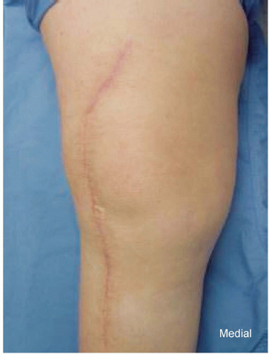 FIGURE 4-1 Large lateral flap used to safely expose the revision knee. |
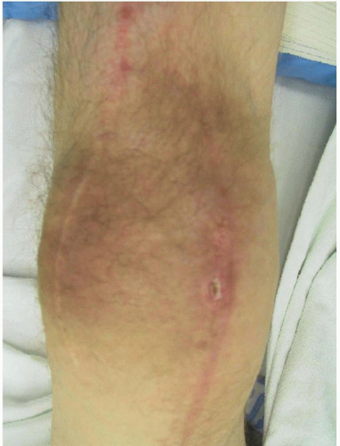 FIGURE 4-2 Medial wound breakdown following use of medial rather than lateral incision. |
Failure to adhere to the rule of using the most lateral incision risks skin necrosis in the bridge of tissue between the two previous incisions. Little comfort should be taken by the operating surgeon by measuring 6 to 8 cm between incisions and then using the most medial incision. This measurement technique may occasionally jeopardize the whole reconstruction if the medial incision is chosen (Fig. 4-2).
Current Range of Motion
The pre-revision range of motion is an important determinant of exposure selection. If the patient has greater than 70 to 80 degrees of range of motion, extensile exposure methods are usually unnecessary. The patellar inversion technique described here can be routinely used. However, if the patient has less than 70 degrees of motion, an “early” quadriceps snip is usually used to safely expose the knee. The quadriceps snip is usually performed approximately 6 cm above the top of the patella. An incision is made from distal medial to proximal lateral at a 45-degree angle within the quadriceps tendon (Fig. 4-3). In extremely tight knees, this incision is carried into the vastus lateralis to facilitate safe exposure.
Previous Surgical Complications
A careful history must be taken to determine if there were any complications related to the extensor mechanism during the primary arthroplasty. Radiographs are helpful in this regard. The presence of suture anchors or staples in the tibial tubercle area should alert the surgeon to be extremely careful in exposing this joint, as the attachment of the previously repaired tendon may be tenuous. In this situation, an “early” quadriceps snip should be done if the surgeon notes that the tissue in the tubercle area tends to peel during routine exposure.
Osteolysis
Related posts:
 Managing Patella Problems in Primary Total Knee Arthroplasty
Cruciate-Retaining Total Knee Arthroplasty
Simultaneous Total Knee Arthroplasty and Femoral Osteotomy
Managing Bone Loss with Metal Augments
Arthrodesis for the Chronically Infected Total Knee Arthroplasty
Unicompartmental Total Knee Arthroplasty With Conventional Instrumentation
Managing Patella Problems in Primary Total Knee Arthroplasty
Cruciate-Retaining Total Knee Arthroplasty
Simultaneous Total Knee Arthroplasty and Femoral Osteotomy
Managing Bone Loss with Metal Augments
Arthrodesis for the Chronically Infected Total Knee Arthroplasty
Unicompartmental Total Knee Arthroplasty With Conventional Instrumentation
Stay updated, free articles. Join our Telegram channel

Full access? Get Clinical Tree


