Fig. 33.1
(a) Macrophotography and (b) specimen radiograph of an epithelioid hemangioma of the distal femur with pathological fracture. The lesion has a hemorrhagic pattern and expands to soft tissue
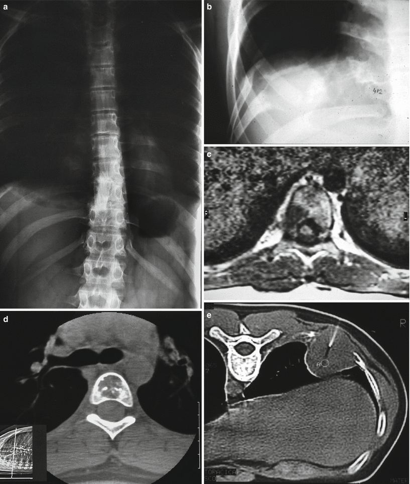
Fig. 33.2
Multifocal EH. (a and b) Radiographs showing lesions in vertebrae and a rib. (c and d) MRI and CT of a vertebral lesion. (e) CT-guided needle biopsy of the rib lesion
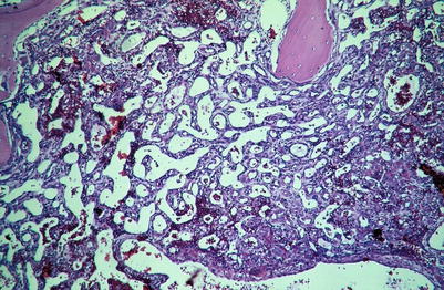
Fig. 33.3
Low-power microscopic view of EH. Endothelium is more flattened at the periphery, left, and more epithelioid towards the center of the lesion
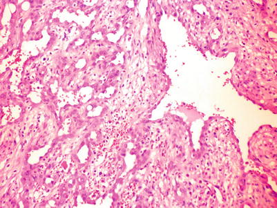
Fig. 33.4
Medium-power view: irregular blood vessels lined by epithelioid endothelial cells
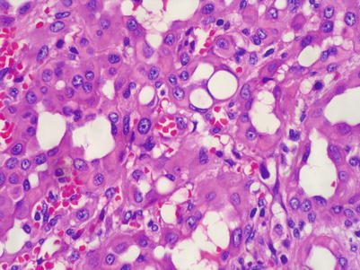
Fig. 33.5
Higher power of a more compacted cellular area
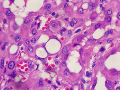
Fig. 33.6


High-power microscopic image. Large, epithelioid cells, with eosinophilic cytoplasm with occasional intracytoplasmic vacuoles and round to oval, sometimes lobulated, nuclei
Related posts:


Stay updated, free articles. Join our Telegram channel
Full access? Get Clinical Tree
