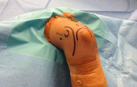
Technique Pearls
Important landmarks should all be drawn out prior to the start of the procedure to aid with positioning and keep the performing surgeon aware of the ulnar nerve. The landmarks include the following: medial and lateral epicondyles, radial head, and olecranon.
Typically the standard 4.0 mm 30° arthroscope is used, but a smaller 2.7 mm arthroscope may be necessary when working on young or smaller patients or in the radiocapitellar joint.
Side-vented inflow cannulas may cause fluid extravasation and should be avoided. An elastic bandage is wrapped around the forearm to further reduce the effect of fluid extravasation.
The joint is distended with approximately 15–25 cc of sterile saline to increase the bone/articular surface to neurovascular structure distance. Of note, this does not significantly change the capsule to neurovascular structure distance.
Applied Technique
Loose body removal
Osteochondritis dissecans – capitellum
ECRB debridement/release for lateral epicondylitis
Radiocapitellar plica
Arthritis/synovitis
Capsular release
Complications
Iatrogenic nerve injury or chondral injury
Collateral ligament injury
Bibliography
1.
Ahmad CS, Vitale MA. Elbow arthroscopy: setup, portal placement, and simple procedures. Instr Course Lect. 2011;60:171–80. Epub 2011/05/11.
2.
Dodson CC, Nho SJ, Williams 3rd RJ, Altchek DW. Elbow arthroscopy. J Am Acad Orthop Surg. 2008;16(10):574–85. Epub 2008/10/04.
7 Elbow Arthroplasty
Take-Home Message
Indications: advanced arthritis/articular incongruity secondary to intra-articular distal humerus fractures with contracture, elderly/inactive patients
Contraindications: prior or current infection, paralysis of upper extremity, inadequate soft tissue sleeve, noncompliance w/ postoperative restrictions
Bryan-Morrey approach and linked, semi-constrained implants preferred
Definition
Surgical reconstruction of the elbow joint replacing the ulnohumeral articulation of the elbow joint with a prosthesis
Indications
Arthritic pain/instability
Osteoarthritis: patients >65 years w/ painful arc of motion and low physical demands
Rheumatoid arthritis: advanced cases (grade III/IV): severe articular cartilage destruction
Post-traumatic arthritis: patients >65 years with low physical demands and significant articular incongruity/instability after an attempted osteosynthesis of an intra-articular fracture/dislocation
Acute trauma
Highly comminuted (or unable to obtain stable fixation) distal humerus fracture in an elderly (>65 years old)/low-demand patient, large, post-traumatic bone defects
Reconstruction after tumor resection
Contraindications
Absolute: active infection, upper extremity paralysis, lack of soft tissue sleeve for coverage, poor patient compliance with postoperative activity limitations
Relative: remote history of elbow infection, neurologic injury involving elbow flexors
Radiographs
AP and lateral elbow radiographs obtained to assess the degree of joint destruction
Preoperative planning to measure humeral bow and medullary canal diameter, and ulnar medullary canal diameter/angulation; if prior total shoulder arthroplasty, must assess for canal length and consider using short-stem humeral component
Cervical spine radiographs (+/− MRI) obtained preoperatively in a patient with rheumatoid arthritis to rule out coexistent cervical spine pathology
CT scan obtained if acute/post trauma to assess degree of articular or metaphyseal comminution and bone defects
Surgical Considerations
Total elbow arthroplasty: prosthetic replacement of the distal humerus and proximal ulna articulations; can use linked (semi-constrained) versus unlinked (resurfacing)
Hemiarthroplasty: distal humerus replaced while preserving ulna/radial head articulations; can be considered in acute trauma setting with intact/repairable collateral ligaments
Interposition arthroplasty: ulnohumeral joint recontouring, release elbow contractures, collateral ligament reconstruction, hinged external fixator applied at end of procedure; considered in post-traumatic arthritis and when the patient is too young or highly active and poor candidate for restrictions required in total elbow arthroplasty
Postoperative Care/Restrictions
Elbow splinted for 24–36 h in full extension followed by open-chain active-assisted range-of-motion exercises
Hemovac used to evacuate hematoma
Restrictions: no pushing or overhead activities ×3 months; no repetitive lifting >5 lb (or single event >10 lbs) weight restriction for lifetime
Complications
Infection, aseptic loosening, short/long term mechanical failure, triceps weakness/avulsion, ulnar nerve injury, periprosthetic fracture, deep venous thrombosis, stiffness/impingement, instability (problematic in unlinked prosthesis)
Bibliography
1.
Celli A, Morrey BF. Total elbow arthroplasty in patients forty years of age or less. J Bone Joint Surg Am. 2009;91(6):1414–8. Epub 2009/06/03.
2.
Day JS, Lau E, Ong KL, Williams GR, Ramsey ML, Kurtz SM. Prevalence and projections of total shoulder and elbow arthroplasty in the United States to 2015. J Shoulder Elbow Surg. 2010;19(8):1115–20. Epub 2010/06/18.
3.
Jenkins PJ, Watts AC, Norwood T, Duckworth AD, Rymaszewski LA, McEachan JE. Total elbow replacement: outcome of 1,146 arthroplasties from the Scottish Arthroplasty Project. Acta Orthop. 2013;84(2):119–23. Epub 2013/03/15.
4.
Kodde IF, Van Riet RP, Eygendaal D. Semiconstrained total elbow arthroplasty for posttraumatic arthritis or deformities of the elbow: a prospective study. J Hand Surg. 2013;38(7):1377–82.
5.
McKee MD, Veillette CJH, Hall JA, Schemitsch EH, Wild LM, McCormack R, et al. A multicenter, prospective, randomized, controlled trial of open reduction-internal fixation versus total elbow arthroplasty for displaced intra-articular distal humeral fractures in elderly patients. J Shoulder Elbow Surg. 2009;18(1):3–12.
6.
Prasad N, Dent C. Outcome of total elbow replacement for distal humeral fractures in the elderly: a comparison of primary surgery and surgery after failed internal fixation or conservative treatment. J Bone Joint Surg B. 2008;90(3):343–8.
7.
Sperling JW, Cofield RH, Schleck CD, Harmsen WS. Total shoulder arthroplasty versus hemiarthroplasty for rheumatoid arthritis of the shoulder: results of 303 consecutive cases. J Shoulder Elbow Surg. 2007;16(6):683–90.
8 Athletic Injuries of the Elbow: MCL Tears
Take-Home Message
Athletic injuries to the elbow can be classified as medial tension injuries, lateral compression injuries, extension overload injuries, and/or tendinopathies
The MCL is composed of three bundles – the anterior, posterior, and oblique bundles. The anterior bundle is the primary stabilizer to valgus stress.
MCL reconstruction with a tendon graft is the primary mode of surgical correction.
Definition
The elbow withstands high forces, especially in athletes that participate in activities that cause repetitive microtrauma, such as baseball players, tennis players, gymnasts, etc. Typically, the injuries fall within medial tension injuries, lateral compression injuries, extension overload injuries, and/or tedonopathies.
Etiology
The causes are widespread and diverse. The etiologies can be divided based on localization of pain.
Medial – medial collateral ligament (MCL) injury, medial epicondylitis
Lateral – osteochondritis dissecans, lateral epicondylitis, radial nerve entrapment, ulnar neuropathy
Anterior – pronator teres syndrome, distal biceps rupture
Posterior – posteromedial impingement, olecranon stress fracture
The MCL is composed of an anterior, posterior, and oblique bundle. It originates at the anteroinferior aspect of the medial humeral epicondyle and inserts onto the sublime tubercle of the ulna.
The anterior bundle is the most important static stabilizer for valgus stability at 20–120° of flexion. Outside this range of motion, the primary stabilizer is the osseous constraint of the olecranon and the trochlea.
Stay updated, free articles. Join our Telegram channel

Full access? Get Clinical Tree


