Dupuytren’s disease and other fibrocontractive disorders

5.2


Dupuytren’s disease and other fibrocontractive disorders


Introduction


Surprisingly, 188 years after Baron Dupuytren first described this condition of the hand, many fundamental details about the disease remain uncertain or unknown. What is known, however, is that it is a disease that affects the fascia in the hand and so it is usually classified as one of the fibrocontractive disorders. It is perhaps a very good example of the contractile power that the fascial elements within a structure can exert when they become “diseased”.


This contractile disorder has received consideration in many textbooks. Highly recommended to the reader is the text written by Tubiana and colleagues in 2000. Despite being 10 years old, this textbook is still the most comprehensive approach to the disease and should be consulted for the detail that cannot be addressed in this short chapter. In contrast, this chapter will draw on and consider in some detail the more recent findings about this condition in an attempt to shed some light on the causes, consequences and possible treatments of this enigmatic disease.


Throughout history the use of the hand has been fundamental to man’s development and many people have praised the hand for its “design” and abilities. One such person was the Englishman, Thomas Traherne (c.1637–1674,) who described the hands as:


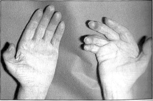
In an individual afflicted by Dupuytren’s disease, there is a usually a progressive decrease in their ability to carry out any tasks they could “imagine or desire” and they are brought to the point of severe frustration and annoyance as the disease progressively and irreversibly develops, as ultimately the result can be a permanent deformity of the hand, with fingers firmly lodged again the palm, incapable of performing their everyday normal functions (Pratt & Byrne 2009). This, it is fully accepted, is the most extreme form of the disease, but when such a state is reached and sometimes even in those people who are suffering the less severe forms of the condition, then the tasks of trying to carry out “anything which reason can imagine or desire” become clearly very difficult and, in the worst case, impossible.


Who is afflicted by this disease?


Dupuytren’s disease is primarily a disease of Northern Europeans, especially those who come from a Celtic or Scandinavian origin. In men, who are more likely to suffer this condition than women, it usually starts during the fourth or fifth decade of life, with a peak incidence at 50 years. In women, the peak incidence is 60–70 years. It does occur in other racial groups throughout the world but at a much lower incidence. The statistics do suggest, however, that there is not a uniformity of occurrence even within a specific Caucasian population. From all the evidence which has so far been collected there appears to be a hereditary component as the disease has a familial trait and autosomal dominance in some cases. Other suggested risk factors include Type 1 diabetes, alcoholism, smoking, antiepileptic drugs, occupations where physical forces are applied to the hand. Some of these factors are more controversial than others. This is just one of the many areas of uncertainty about the etiology of the disease which remain to be resolved.


A further complication which makes the disease even more complicated to understand is that not all individuals show the same rate of progression: usually, the later it starts the less aggressive it proves to be. The disease has an association with other fibrocontractive disorders such as Peyronie’s and Ledderhose’s disease (see below).


The basic problems of Dupuytren’s disease


The beautiful delicacy, complexity and dexterity of the hand is brought about by the highly coordinated interaction of nerves, muscles, tendons, and bones, which just like everywhere else in the body are connected together with fascial elements. In the hand these elements are arranged in a very complex way and none more so than those that are found directly under the palmar skin. These elements are somewhat unusual since they form a thickened sheet of connective tissue, which is termed the palmar aponeurosis. This thickened layer of connective tissue is thought to provide physical protection for the flexor tendons which lie directly underneath it on their route to the fingers, so ensuring that despite stresses and loads placed upon the palm, the fingers can be manipulated. This ensures that these tendons are able to carry out their delicate and complex tasks free from constraints resulting from externally applied forces.


It is this connective tissue structure – the aponeurosis – which is considered to be central to the palmar problems of Dupuytren’s disease. There is great speculation as to why this structure is affected by the disease and what factor(s) initiate the disease. There has also been speculation about what factors cause the condition to progress once it starts. It could be that these factors are the same or they could be different. Before speculating about these factors and what they could potentially do, it may be useful to give a brief outline of the condition, with special reference to the involvement of the fascial elements in the hand.


Basic anatomy of Dupuytren’s disease


Many studies, from the perspectives of both basic anatomy (gross and microscopic) and clinical medicine, have established that the first palmar signs of Dupuytren’s disease are the development of “skin pits”, especially at the position of the distal palmar crease. These pits are caused by changes in the underlying vertical fibers of the aponeurosis attached to the overlying subcutaneous fascia of the skin. They are usually slow to develop and are due to progressive “stimulation” of the existing vertical connective tissue elements within the aponeurosis.


It should be stressed that the vertical fibers are fewer in number than the more extensive longitudinal and transverse arrangement of fibers within the structure, which provide skin anchorage when the palmar skin is subjected to shearing forces. So, the power of the vertical fibers to exert a force on the overlying skin is relatively low. However, once stimulated, these fibers do transmit a force and pits result from the contraction pulling against the more distensible skin rather than their anchorage points in the palmar aponeurosis.


This suggests that from the very beginning of the disease, the palmar fascia has changed from a highly complex, organized, passive protective structure to one which has developed the capacity to be contractile. Despite speculation, there is still no clear evidence as to why the vertical transmission of the contractile force that forms the pits should first occur. However, whatever the causes, the presence of such fibres as demonstrated in the micro dissection studies of McGrouther in 1982 is not in any doubt. McGrouther clearly demonstrated the vertical connections of the palmar aponeurosis to the skin at the site of the distal palmar crease.


Palmar nodules


The disease can then enter a very variable period. In some people, the disease does not progress from this stage or progresses very slowly. In others, there is proliferation of cells on the surface of the palmar aponeurosis and over time, as the number of cells increases, their mass is such that they can form a distinct, palpable and, eventually, externally visible, nodule full of cells (palmar nodules). It should be stressed that palmar nodules are superficial to the palmar aponeurosis and not an integral part of it. They exist between the palmar aponeurosis and the fascia of the overlying skin to which they can be adherent.


The proliferation of the cells in palmar nodules can be so pronounced that in times past when they were examined histologically they were sometimes diagnosed as malignant tumors (such as a sarcoma), resulting in amputation. Today, greater knowledge of the disease means such radical surgery is avoided. The proliferating cells are found in a meshwork of connective tissues of such a density that when they are bisected using a scalpel blade the term “gritty” has been used to give an indication of their great density, which is unlike that of the unaffected palmar fascia.


The condition gives rise to new connective tissue fibers and also new cells in a location where there should be no unnecessary connective tissue or additional cells. Biochemical studies have established that the newly formed collagen is a mixture of type III and type I, with collagen II being the predominant type associated with the nodules. The cells within the nodules have some of the functional characteristics of fibroblasts – synthesizing this new collagen – and ultrastructural similarities to some aspects of smooth muscle cells – giving rise to the contraction seen in the disease (Fig. 5.2.1).


The cells which develop within the palmar nodules have some rather unusual morphological characteristics when viewed using both optical and electron microscopy. They exhibit nuclear pleiomorphism using both techniques. With the greater resolution of the electron microscope, their cytoplasm contains myofilaments and there are also associated dense bodies. It is interesting to note that in 1972 Gabbiani and Majno used these ultrastructural appearances in such palmar nodules to develop further their ideas as to the existence and possible role(s) of myofibroblasts.


As the disease develops further and more nodular cells are formed, the greater contractile force generated by these myofibroblasts is transmitted to the rest of the aponeurosis along the longitudinal fibers. These consequently thicken and exert force on the proximal metacarpophalangeal joint (MP joint). Further disease progression results in palmar “cords” being formed, which may then spread their contractile effects to the MP joint and so cause deformity.

Stay updated, free articles. Join our Telegram channel

Aug 24, 2016 | Posted by in ORTHOPEDIC | Comments Off on Dupuytren’s disease and other fibrocontractive disorders

Full access? Get Clinical Tree

Get Clinical Tree app for offline access