Distal Radius Osteoarticular Allograft
Miguel A. Ayerza
Luis A. Aponte-Tinao
D. Luis Muscolo
Osteoarticular allografts of the distal part of the radius replace an articular surface for which prostheses are notreadily available. The reconstruction of distal radius after tumor resections, preserving wrist stability and function, is limited. Osteoarticular allografts are utilized for reconstruction of one side of the joint after tumor resection. Osteoarticular allografts do not sacrifice the side of the joint that is not compromised by the tumor, they can replace articular surfaces for which prostheses are not readily available, such as the distal radius, and can be attached to soft tissues.
INDICATIONS
The procedure is appropriate for the treatment of a massive osteoarticular defect after tumor resection or massive traumatic bone losses of the distal radius (Figs. 36.1 and 36.2).
At least one of the two major neurovascular bundles, the radial or the ulnar artery, must be free of tumor.
The distal ulnar epiphysis must remain intact with its ligamentous insertions.
Appropriate soft tissue coverage.
CONTRAINDICATIONS
Patients with sarcomas in whom preoperative imaging studies demonstrate evidence of intra-articular compromise of the tumor.
Inadequate host soft tissue to reconstruct the defect.
Patients with cartilage degenerative disease of the wrist.
Absence of the distal ulna or ligament insertions.
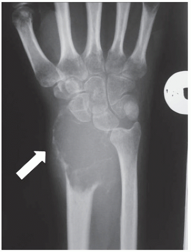 FIGURE 36.1 Anteroposterior radiograph of the distal forearm showing a giant cell tumor in the distal radius. |
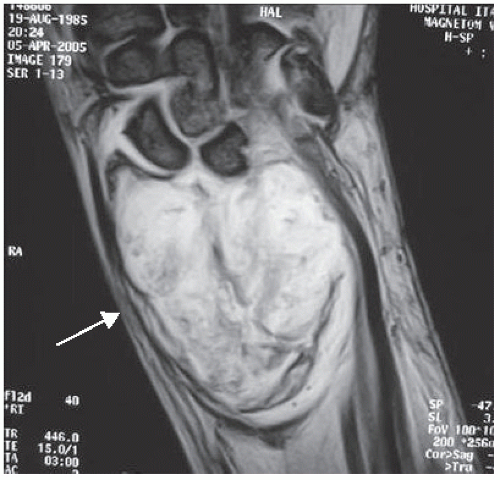 FIGURE 36.2 Coronal magnetic resonance image that illustrates the extent of the tumor. |
PREOPERATIVE PREPARATION: ALLOGRAFT SELECTION
Fresh-frozen allografts are obtained and stored according to a technique that has been previously described (1). Poor anatomical matching of both size and shape between the host defect and the graft can significantly alter joint kinematics and load distribution, leading to bone resorption or joint degeneration. To improve accuracy in size matching between the donor and the host, we developed measurable parameters based on CT scans of the distal radius. In the axial view of the distal radius, we measure the maximum total width and anteroposterior width of the radius. The allograft is selected on the basis of a comparison of these measures with those of the host.
TECHNIQUE
All operations are performed in a clean-air enclosure with vertical airflow and usually with regional anesthesia. The patient is placed on the operating table in the supine position and an arm table is added with the upper limb placed in position. A long dorsal incision is made following the third metacarpal, and the extensor tendons are exposed (Fig. 36.3). Once these tendons are dissected, the wrist joint is identified and a dorsal carpal arthrotomy is performed (Figs. 36.4 and 36.5). The biopsy track is left in continuity with the specimen. The distal radioulnar joint is identified and sectioned near the radius insertion to ensure that there is enough host tissue to reconstruct these ligaments to the allograft. Palmar radio scaphoid ligaments are sectioned in the same fashion in order to preserve enough host tissue for reconstruction. If there is an extraosseous tumor component, a cuff of normal muscle (i.e., pronator quadrates) must be included in the resection. An osteotomy of the radius proximal to the tumor is performed at the appropriate location as determined on the basis of the preoperative imaging studies. All remaining soft tissues at the level of the transection are cleared. After the posterior and medial structures have been protected and retracted, the osteotomy is performed perpendicular to the long axis of the radius (Fig. 36.6). Following the osteotomy, the distal radius is pulled dorsally in order to expose the soft tissue attachments of the palmar space (Fig. 36.7). These palmar soft tissue structures are transected and the distal radius is then passed off the operative field (Fig. 36.8).
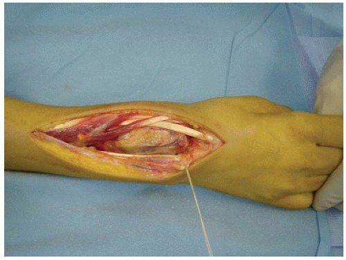 FIGURE 36.3 Intraoperative photograph that shows the extended dorsal approach that is employed. |
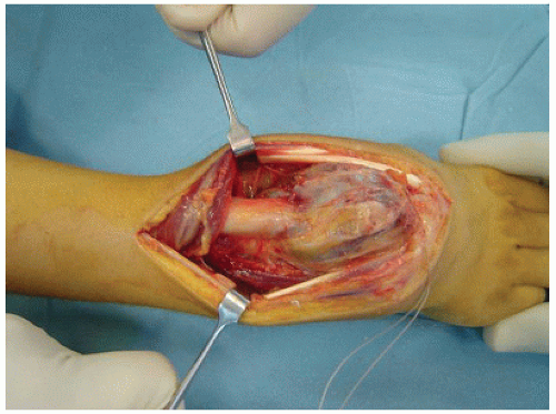 FIGURE 36.4 Intraoperative photograph showing wide exposure of the distal radius before the arthrotomy is performed. |
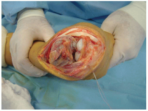 FIGURE 36.5 Intraoperative photograph obtained when the dorsal carpal arthrotomy is performed. |


