37
Distal Phalangeal Fractures
Kevin D. Plancher
History and Clinical Presentation
A 35 year old carpenter was on the job using a table saw. He forgot to engage the safety and cut the tip of his finger. He presents to the emergency room with exposed bone at the tip of his finger after a transverse cut to his fingertip.
Physical Examination
There is no soft tissue covering the end of the bone. Approximately 40% of the nail and nail matrix are present.
Diagnostic Studies
Anteroposterior (AP), lateral, and oblique radiographs of the injured digit were obtained. The finger was assessed for foreign objects, joint dislocation, subluxation, and fractures.
Differential Diagnosis
Distal phalanx fracture
Vascular injury
Nerve injury
Mallet fracture
Distal phalanx (P3) dislocations
PEARLS
- Expect patients 50% of the time to have cold intolerance, hypersensitivity, and paresthesias.
- Sew these flaps with chromic or catgut sutures. Suture to the nail should be absorbable to avoid painful removal in the office.
PITFALLS
- Attention to detail in lifting the V-Y flap in the subcutaneous tissue will provide survival of the tissues.
- Adequate incision and drainage with antibiotics will avoid infection in most cases.
Diagnosis
Open Distal Phalanx Fracture with Soft Tissue Loss
The type of injury, the functional goal, and possible complications often determine the treatment of fingertip injuries. Factors to consider include finger sensitivity, a nontender finger, maximum length, nail appearance, normal joint movement, and cosmesis. On initial diagnosis, the viability of the tissues should be assessed to determine which tissue is unlikely to survive. It must also be determined what can be sacrificed and what must be preserved to maintain function.
Transverse amputations are the easiest to repair. Treatment is determined by the coverage of the bone. The amount of nail following injury is also important in determining treatment. Depending on the level of amputation, blood vessel and nerve status must also be determined. In oblique dorsal amputations, if the nail is only partially injured, an attempt must be made to preserve it. The condition of the nail matrix is also important. Oblique palmar amputations are difficult to treat. Thenar flaps are an option. The goal of treatment in a palmar amputation is to provide a reconstructed pulp, which is sensitive and well cushioned.
Surgical Treatment
For this patient the volar V-Y flap or Atasoy technique was used to provide coverage of exposed bone with adequate padding. The patient was most concerned with fingertip tenderness and function of the finger.
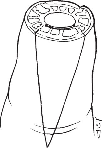
The finger was prepared and a lidocaine metacarpal block supplemented with bupivacaine provided anesthesia. A Penrose drain was placed on the digit to act as a tourniquet. The wound was irrigated and surgically cleaned, and debridement of the skin edges was performed. A pattern of the defect was transferred onto the palmar skin proximal to the defect. The skin pattern was made ∼1 mm larger. The palmar skin incisions were marked from the lateral edges of the defect site to enclose the skin pattern and continued proximally and obliquely to meet in the midline of the finger at the distal crease (Fig. 37–1). The skin was incised through the dermis sharply and the flap was dissected from the periosteum and the flexor sheath. The Grayson’s and Cleland’s ligaments that surround the neurovascular bundles and connect the flap to surrounding tissues were bluntly dissected and divided with dissection scissors (Fig. 37–2). The flap was mobilized to allow advancement of the flap without tension (Fig. 37–3). Hemostasis was maintained with a bipolar cautery.
The flap was then sutured to the nail with nonabsorbable sutures (Fig. 37–4). The apex of the V was closed with interrupted sutures, which were kept close to the skin edge to avoid interference with the vascular supply. The tourniquet was deflated and blood supply to the flap was verified. The finger was treated with Xeroform, a light dressing, and a protective tip splint. The patient was instructed to keep the finger elevated for 2 days. The dressing was changed 5 days postoperatively and the patient was prescribed active exercises of the proximal interphalangeal (PIP) joint. Sutures were removed at 14 days postoperatively and the patient was instructed to keep the protective splint on for 4 to 6 weeks postoperatively.