, George C. Babis2 and Kalliopi Lampropoulou-Adamidou3
(1)
Orthopaedic Department Medical School, National and Kapodistrian University, Athens, Greece
(2)
2nd Orthopaedic Department Medical School, National and Kapodistrian University Nea Ionia General Hospital “Konstantopoulion”, Athens, Greece
(3)
3rd Orthopaedic Department, National and Kapodistrian University KAT General Hospital, Athens, Greece
Abstract
Total hip replacement in low and high dislocation is a difficult operation because of the anatomical features of these hips. Previous unsuccessful treatments increase considerably the surgical difficulties. Regardless the method and implants used for the reconstruction of these hips, wide exposure is mandatory and it is achieved when greater trochanter is osteotomised. Also, proximal shortening of the femur, when needed, facilitates the reconstruction of these difficult cases.
Total hip replacement (THR) in low and high dislocation is a difficult operation because of the anatomical features of these hips. Previous unsuccessful treatments increase considerably the surgical difficulties. Regardless the method and implants used for the reconstruction of these hips, wide exposure is mandatory and it is achieved when greater trochanter is osteotomised. Also, proximal shortening of the femur, when needed, facilitates the reconstruction of these difficult cases.
Clinical and radiological results in the majority of these cases are less satisfactory and reoperations more often. Examples are following (all patients were females).
Case 1
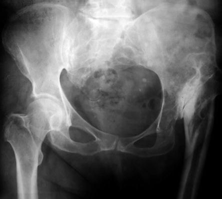
A 48-year-old patient with severe bilateral secondary osteoarthritis (OA) due to low dislocation. Both hips were stiff and almost fixed in 40° flexion. The patient at the age of 33 had in an other institution an intertrochanteric osteotomy on the left hip (Figs. 9.1, 9.2 and 9.3).
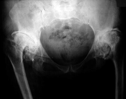
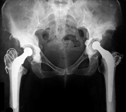
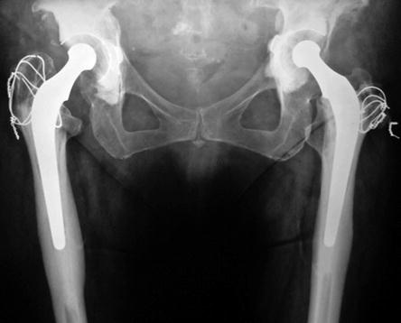
Fig. 9.1
Preoperative radiograph
Fig. 9.2
One year after low-friction arthroplasty (LFA) of both hips. The patient gained compensatory range of motion and almost full level of activity
Fig. 9.3
Twenty years after surgery. The good result remains
Case 2
A 37-year-old patient with low dislocation of both hips. In 1952, at the age of 4, she had an operation of unknown type on the left hip followed by prolonged immobilisation in hip spica for 2.5 years according to patient’s statement (Figs. 9.4, 9.5, 9.6 and 9.7).
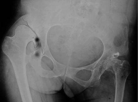
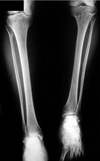
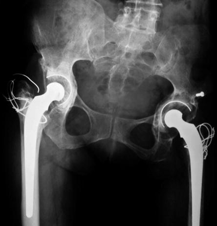
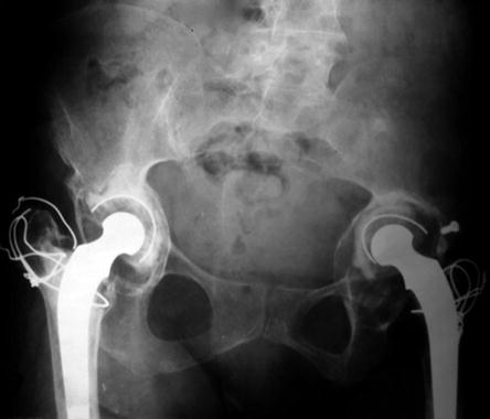
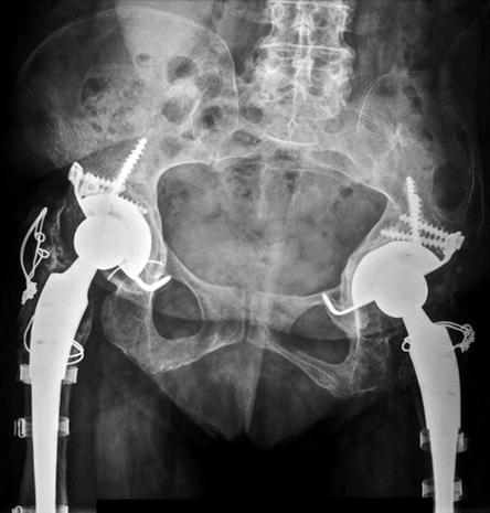
Fig. 9.4
Preoperative radiographs. The left leg was 10 cm shorter probably due to the previous operation in childhood
Fig. 9.5
Ten years after bilateral LFA. The cotyloplasty technique was used in both hips with offset-bore cups. In the femoral side, the CDH stems of Charnley were used. Seven centimetre shortening remained on the left leg
Fig. 9.6
Nineteen years postoperatively both acetabular components were loose and needed revision
Fig. 9.7
Seven years after revision of both components on both hips. Patient has no pain but a limited level of activity. For the acetabuli, cementless Hilock cups (Symbios) were used, and for the femoral side, cementless modular revision stems (Profemur, Wright)
Case 3
Thirty-four-year-old patient with high dislocation on the right hip and dysplasia on the left. According to her medical record, at the age of 4 (1957), she had Schanz osteotomies on both hips (Figs. 9.8, 9.9 and 9.10).
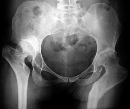
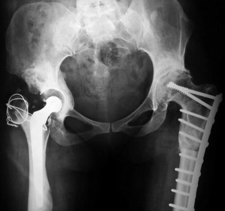
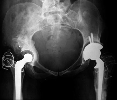
Fig. 9.8
Preoperative radiograph. Reconstruction of both hips was performed as follows: (1) THR on the right hip by shortening of the femur at the level of the neck. (2) Corrective osteotomy of the left hip. (3) THR on the left hip (see also Fig. 6.7)
Fig. 9.9
Radiograph after LFA with cotyloplasty and offset-bore acetabular component on the right hip and corrective osteotomy on the left. After 2 years, a hybrid THR was followed on the left hip
Fig. 9.10
Latest follow-up radiograph when the patient was 45 years old. She died 2 years later from breast cancer
Case 4
This patient with high dislocation of the left hip was examined by us when she was 62 years old. A Schanz osteotomy was performed in 1963 when she was 35 years old (Figs. 9.11 and 9.12).