, George C. Babis2 and Kalliopi Lampropoulou-Adamidou3
(1)
Orthopaedic Department Medical School, National and Kapodistrian University, Athens, Greece
(2)
2nd Orthopaedic Department Medical School, National and Kapodistrian University Nea Ionia General Hospital “Konstantopoulion”, Athens, Greece
(3)
3rd Orthopaedic Department, National and Kapodistrian University KAT General Hospital, Athens, Greece
Abstract
The Chapter includes the complications and the results of total hip replacement in patients with congenital hip disease presented in the literature and in the authors’ cases.
8.1 Complications
It has been reported that complications associated with total hip replacement (THR) in patients with congenital hip disease (CHD) are higher, according to the severity of the disease, when compared to those in patients with primary osteoarthritis [1]. These complications include nerve palsies and dislocations .
Sciatic and femoral nerves are the most vulnerable nerves to be damaged during THR [2, 3]. The overall incidence of nerve palsy after THR has been reported to be 0.8–3.7 % [4], while in a large number of patients with CHD, Schmalzried et al. found prevalence of nerve palsies 5.2 % after THR [5]. Several authors have associated nerve palsies with leg-lengthening in cases with CHD. Farrell et al. [6] reported that the preoperative diagnosis of CHD and the leg-lengthening were associated significantly with the development of postoperative nerve palsies (p = 0.0004 and p < 0.01, respectively). There is a controversy regarding the safe amount of leg-lengthening among different authors. Garvin et al. considered the 2 cm [7], while Edwards et al. the 4 cm [8]. However, in 13 cases of our series where leg-lengthening surpassed 5 cm (5–7), no neurological complications were observed [9]. Also, Eggli et al. [10], in a series of 508 THRs for CHD, found no statistical correlation between the amount of leg-lengthening and the incidence of nerve damage (p = 0.47), while they found significant correlation between nerve palsy and intraoperative difficulty (p = 0.041) such as previous surgery, severe deformity, a defect of the acetabular roof or considerable flexion deformity.
Higher rates of postoperative dislocation, in patients with CHD, are also reported. The Norwegian Arthroplasty Registry [11] found that THRs for CHD have 5.6 times higher risk of revision due to dislocation when compared with THRs for primary osteoarthritis. The risk of postoperative dislocation has been reported to increase with trochanteric non-union , high hip centre , medialisation of the cup [12], small femoral head size [13, 14] and large acetabular component outer diameter [14].
Hypoplastic or deformed femoral diaphysis in hips with CHD leads to increased risk of intraoperative femoral fracture which is reported to range from 5 to 22 % [15], and therefore care must be taken when preparing the femoral canal to avoid cortical perforation (see Chap. 7). The postoperative infection rate is reported as higher as ten times in patients with CHD when compared to other diagnoses. This could be due to the complexity and duration of these operations, the large exposure and extensive dissection, soft tissue stripping and the frequent use of bone grafts [16].
We have registered the complications of 223 THRs performed in 162 patients with osteoarthritis secondary to CHD: 76 dysplastic hips, 69 hips with low dislocation and 84 hips with high dislocation [17]. In the dysplastic group, there was one postoperative femoral nerve palsy that resolved within 1 year. One patient with a low dislocation presented with palsy of both the peroneal and the femoral nerve on the fifteenth postoperative day, possibly as a result of a perineural hematoma. The femoral nerve palsy resolved completely within 6 months, but the peroneal nerve palsy resulted in a permanent incomplete drop foot . In the group of hips with a high dislocation, there was one peroneal nerve and one femoral nerve palsy. Both fully resolved within 6 months. Postoperative dislocations occurred in eight hips. In three hips, the acetabular component was revised because of incorrect orientation. Five dislocations occurred at 7–52 days following hybrid total hip arthroplasties (during the initial period of application of this method); one occurred in a hip with a low dislocation and four in hips with a high dislocation. Four of these dislocations were treated with closed reduction and one with open reduction. No recurrence of any of the eight dislocations was recorded. Femoral fracture occurred in a hip with low dislocation 22 years postoperatively and treated with internal fixation . One dysplastic hip, three hips with low dislocation and three with high dislocation developed deep infection.
To evaluate the complications associated with trochanteric osteotomy, we studied 192 THRs in 140 patients with CHD: 34 dysplastic hips, 93 with low dislocation and 65 with high dislocation [18]. The non-union rate of the osteotomy was 3 % (5 hips). Fibrous union was noticed in 29 hips (15 %) resulting in a mild Trendelenburg gait postoperatively. The rate of union had a statistically significant relationship with the position of reattachment of the trochanter, which depended greatly on the preoperative diagnosis. Acetabular and femoral loosening had a statistically significant relationship with defective union (non-union and fibrous union) and the position of reattachment of the trochanter. Different authors have reported that trochanteric osteotomy increases the risk of heterotopic ossification [19–22]. However, Brooker et al. [23] stated that unless there was ankylosis (Brooker IV), the functional result is not affected. In our series, Brooker grade IV heterotopic ossification was seen in only three hips (2 %) resulting in complete ankylosis in one hip (Fig. 8.1) and gross limitation of movement in the other two. We suggest that in routine cases of THR, trochanteric osteotomy may be optional, but it remains a useful technique in cases with CHD, especially those with low and high dislocation.
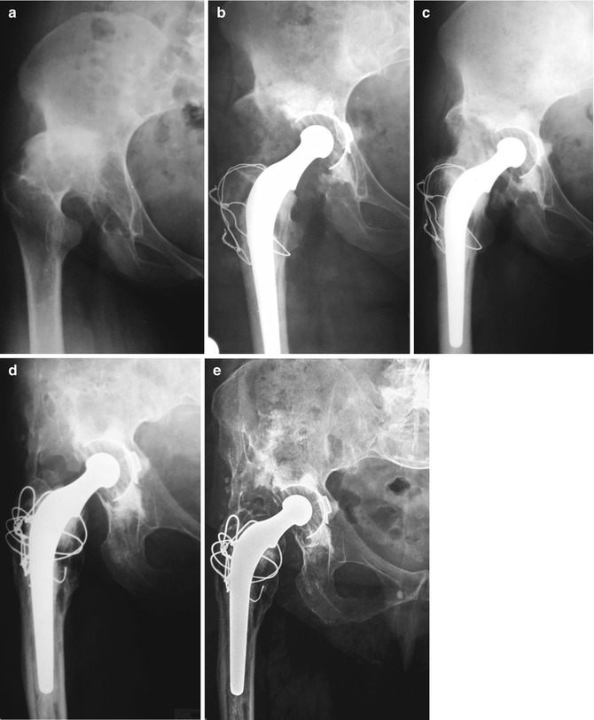
Fig. 8.1
(a) Radiograph of a 43-year-old female patient with low dislocation of B2 subtype on the right hip. (b) Two months after low-friction arthroplasty. (c) Radiograph 1 year after surgery with complete ankylosis due to Brooker IV heterotopic ossification. (d) Radiograph after excision of the largest part of the heterotopic ossification. Patient regained 70° of flexion, 15° abduction, 10° adduction and 15° of external rotation. (e) Final radiograph taken 34 years after primary surgery
With proper surgical technique and postoperative care , complications, especially nerve damage and postoperative dislocation, can be minimised. Nerve damage can be avoided by cautious handling of the various retractors intraoperatively and, in high or low dislocations, by the placement of both the hip and the knee in flexion for 3–4 days after the operation. Postoperative dislocation can be minimised by proper orientation of the cup (inclination of 30–45° in the frontal plane and 10–15° of anteversion).
8.2 Results
While reporting results of total hip replacement (THR), long-term follow-up is of paramount importance. Short-term results are unreliable.
The reported results of THR in patients with congenital hip disease (CHD) are not easily comparable (Table 8.1) [2, 7, 17, 24–40]. The material in the majority of reports is not homogenous, since they include different types of the disease, duration of follow-up, implants and techniques used and statistical analysis. In five relatively homogenous series of high dislocated hips that we are aware, it is reported 25 % failure rate in a group of 87 hips at an average 10 years (5–16) of follow-up [26], 14.7 % in a smaller group of 34 hips at an average 9.4 years (5.6–14) of follow-up [27], 12.7 % in a group of 118 hips at an average 12.8 years (10–26) of follow-up [33], 17 % in a group of 116 hips at an average 9.7 years (6–14) of follow-up [36] and 20 % in a group of 20 hips at an average 10.2 years (5–20) of follow-up [40].
Table 8.1
Mid- and long-term reported results of THR
Year of publication | Authors | No of hips | Range of follow-up (years) | CHD type (no of hips) | Failure rate (%) |
|---|---|---|---|---|---|
1979 | Crowe et al. [2] | 31 | 2–6 | Severe dysplasia, dislocation | 9.7 |
1988 | Linde et al. [24] | 129 | Not referred | Congenital dislocation and subluxation | 11 |
1991 | Fredin et al. [25] | 21 | 5–11 | Crowe type IV | 23.8 |
1991 | Garvin et al. [7] | 23 | 8–16.5 | Severe dysplasia, dislocation | 26.0 |
1991 | Kavanaugh et al. [26] | 87 | 5–16 | Complete dislocation | 25 |
1993 | Anwar et al. [27] | 34 | 5.6–14 | Congenital dislocation | 14.7 |
1993 | Paavilainen et al. [28] | 67 | 3–5 | Severe dysplasia, dislocation | 44.8 |
1995 | Morscher [29] | 71 | 1–9 | Dysplasia, dislocation | 12.7 |
1996 | MacKenzie et al. [30] | 59 | 10–21 | Crowe type II, III, IV | 13.6 |
1997 | Nagano et al. [31] | 34 | 15–23.3 | Congenital hip dysplasia | 11.8 |
1997 | Numair et al. [32] | 232 | 3.1–22.8 | Crowe type I, II, III (136) | 9a |
3b | |||||
Type IV (46) | 15a | ||||
2b | |||||
2001 | Kerboull et al. [33] | 118 | 10–26 | Crowe type IV | 12.7 |
2003 | Ito et al. [34] | 81 | 8–15 | Hartofilakidis type I (70), II (7), III (4) | 2.5 |
2004 | Hartofilakidis and Karachalios [17] | 223 | 7–26 | Hartofilakidis type I (76) | 21 |
Type II (69) | 28 | ||||
Type III (84)
Related posts:Stay updated, free articles. Join our Telegram channel
Full access? Get Clinical Tree
 Get Clinical Tree app for offline access

|
