Fig. 5.1
Histology of Sectioned Ovine Retrieval at 26 Weeks Demonstrating Bone-Through Growth into the Cage Peg
Classification of RTSA Implant Designs
RTSA Design Classification System
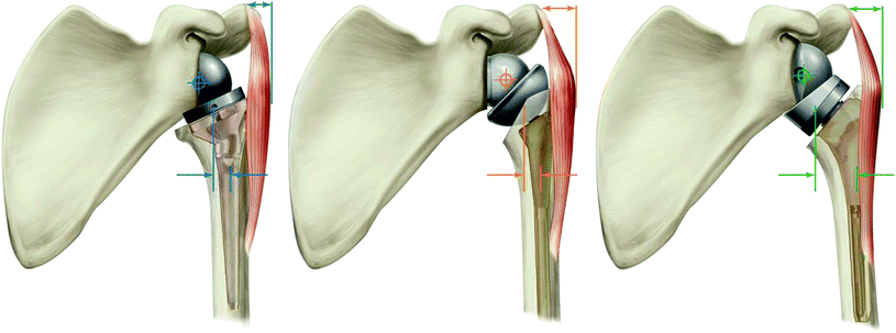
The proliferation of RTSA prosthesis designs and design philosophies has necessitated the need for a classification system to better understand differences between prostheses, to clarify differences in surgical techniques, and to better enable a comparison of clinical outcomes. We contend that the 2 defining characteristics that distinguish RTSA prosthesis designs are the position of the CoR relative to the native glenoid (be it, medial or lateral, as that influences muscle moment arms and torque on the glenoid fixation surface) and position of the humerus (be it, medial/intramedullary or lateral/extramedullary, as that influences muscle tensioning and deltoid wrapping). These different methods to modify the CoR and humerus led us to propose the following RTSA design classification system [21] (Fig. 5.2).


Fig. 5.2
RTSA design classification system; from left to right: medial glenoid/medial humerus (MGMH), lateral glenoid/medial humerus (LGMH), and medial glenoid/lateral humerus (MGLH) RTSA prosthesis designs. Depicted on each is the center of rotation, lateral distance from the center of humeral stem to deepest portion of the humeral cup, and the resulting humeral lateralization
Medial Glenoid/Medial Humerus (MGMH) Design
In the MGMH design category, the glenoid component CoR is positioned medially on or near the native glenoid and the humeral component is intramedullary so that the humerus is positioned below the glenosphere in a relatively medial configuration. Characteristics of this design include a low glenoid loosening rate [13, 15, 18], a demonstrated history of restoring active forward flexion and abduction (due to the large deltoid abductor moment arm) [13, 15–17, 25, 26, 33, 34, 41, 42], a high scapular notching rate [15–17, 25, 26, 33–35, 41–43], poor improvements in active internal and external rotation (due to the shortened rotator cuff muscles) [13, 15–17, 22, 25, 26, 33, 34, 41, 42], and a requirement to repair the subscapularis to maintain stability [44].
Lateral Glenoid/Medial Humerus (LGMH) Designs
In the LGMH design category, the glenoid component CoR is positioned lateral to the native glenoid either by use of a thicker glenosphere (relative to its diameter) or by use of bone graft behind the baseplate and the humeral component is intramedullary so that the humerus is positioned below the glenosphere. However, because the glenoid component is lateralized, the humeral component is positioned in a more lateral configuration relative to the MGMH design. Characteristics of this design include a slightly higher glenoid loosening rate (relative to the MGMH designs), a significantly reduced scapular notching rate, and better improvements in active internal and external rotation (due to better tensioning of the rotator cuff muscles) [45–48]. Additionally, subscapularis repair may not be required to maintain stability as the more lateral humeral position better restores rotator cuff muscle tension and deltoid, both of which contribute to stability [22, 49].
Medial Glenoid/Lateral Humerus (MGLH) Designs
In the MGLH design category, the glenoid component CoR is positioned medially on or near the native glenoid and the humeral component is extramedullary, with the humeral liner resting on top of an anatomic humeral head osteotomy in a relatively lateral position. Characteristics of this design include a low glenoid loosening rate [37–39, 50, 51], a reduced scapular notching rate [19, 20, 24, 27], a demonstrated history of restoring active forward flexion and abduction (due to the large deltoid abductor moment arm) [52], and better improvements in active internal and external rotation (due to better tensioning of the rotator cuff muscles) [22, 52]. Additionally, subscapularis repair may not be required to maintain stability as the more lateral humeral position better restores rotator cuff muscle tension and deltoid, both of which contribute to stability [22, 53].
This RTSA classification system can be expanded to include different or more detailed changes in the position of the CoR relative to the native glenoid (e.g., inferior, superior, posterior, and inferior–posterior) or position of the humerus (e.g., anatomic lateralization, and posterior shift) as RTSA prosthesis designs continue to evolve. Three refining examples are as follows:
1.
A lateral glenoid/lateral humerus (LGLH) design would have a CoR positioned lateral to the native glenoid either by use of a thicker glenosphere (relative to its diameter) or by use of bone graft behind the baseplate and an extramedullary humeral component. As both the glenoid and humeral components are lateralized relative to MGMH designs, the position of the humerus with the LGLH designs is more lateral than both LGMH and MGLH designs.
2.
It is possible to medialize the CoR more than proposed with the Grammont design using a glenosphere whose thickness is less than its radius; doing so further increases the deltoid abductor moment arms. This inset CoR glenosphere design could be termed as extra medial glenoid and could be used with intramedullary or extramedullary humeral designs.
3.
It is possible to posteriorly shift the humerus by offsetting the humeral tray of an extramedullary design; doing so increases the length of the external rotation moment arms and better tension the posterior rotator cuff [54, 55]. This design could be termed as medial glenoid/posterior–lateral humerus.
Impact of Reverse Shoulder Prosthesis Design on Muscle Characteristics
Impact of RTSA Design and Implantation Techniques on CoR and Position of the Humerus
Inverting the concavities with RTSA causes an inferior-medial shift in both the CoR and position of the humerus relative to the anatomic shoulder. The magnitude of this inferior–medial shift is influenced by both prosthesis design and implantation techniques. A recent computer study quantified how CoR and humeral position change with different RTSA designs and with different glenoid (no tilt vs. 15° inferior tilt and with or without bone graft) and humeral (0°, 20°, and 40° humeral retroversion) implantation techniques [22]. As described in Table 5.1, each design and implantation technique results in different amounts of medial and inferior shifts in both the CoR and humerus, where the combination of medial and inferior shifts alter the angle of shift [22].
Table 5.1
Change in CoR and humerus position for each reverse shoulder design and implantation technique relative to normal anatomic shoulder [22]
Medial shift in CoR (mm) | Inferior shift in CoR (mm) | Angular shift in COR (inferior/medial) | Medial shift in humerus (mm) | Inferior shift in humerus (mm) | Angular shift in humerus (inferior/medial) | |
|---|---|---|---|---|---|---|
36 Grammont, 0° tilt, 20° retro | 28.3 | 8.0 | 15.8° | 21.5 | 30.2 | 54.6° |
36 Grammont, 15° tilt, 20° retro | 31.0 | 7.7 | 13.9° | 24.2 | 29.9 | 51.0° |
36 Grammont, 0° tilt, 0° retro | 28.3 | 8.0 | 15.8° | 19.9 | 30.1 | 56.5° |
36 Grammont, 0° tilt, 40° retro | 28.3 | 8.0 | 15.8° | 23.6 | 30.1 | 51.9° |
36 Grammont, graft, 0° tilt, 0° retro | 19.2 | 8.0 | 22.6° | 10.8 | 30.1 | 70.3° |
36 Grammont, graft, 0° tilt, 20° retro | 19.2 | 8.0 | 22.6° | 12.4 | 30.1 | 67.6° |
36 Grammont, graft, 0° tilt, 40° retro | 19.2 | 8.0 | 22.6° | 14.5 | 30.1 | 64.3° |
36 Grammont, graft, 15° tilt, 20° retro | 21.9 | 10.2 | 25.0° | 15.1 | 32.4 | 65.0° |
32 RSP, 0° tilt, 20° retro | 20.0 | 6.9 | 19.0° | 11.7 | 25.3 | 65.2° |
38 Equinoxe, 0° tilt, 20° retro | 27.1 | 4.5 | 9.4° | 9.1 | 34.8 | 75.3° |
Impact of RTSA Design and Implantation Techniques on Muscle Tensioning and Deltoid Wrapping
The magnitude of inferior–medial shifts in the CoR and humerus has important biomechanical implications on muscle origin-to-insertion distances and how these muscles cross-over and wrap around the humerus. For example, at low levels of elevation, the middle deltoid wraps around the greater tuberosity to compress the humeral head into the glenoid [56]. While this compressive force is small relative to that generated by the rotator cuff, preserving deltoid wrapping may be an important contributor to stability with RTSA [22, 56]. Thus, differences in humeral position may explain why some RTSA designs better restore active internal and external rotation than the Grammont design and may also describe why some RTSA designs do not have an increased risk of instability when the subscapularis is not repaired [22, 49, 53].
The aforementioned computer study [22] also quantified how muscle tension and deltoid wrapping change with different RTSA designs and different glenoid and humeral implantation techniques. As described in Table 5.2, each of these different designs and implantation techniques result in reduced deltoid wrapping, deltoid elongation, and rotator cuff muscle shortening. Designs and implantation techniques that result in less humeral medialization were associated with more anatomic deltoid wrapping and more anatomic rotator cuff tensioning. With regard to RTSA design classification, the MGMH (e.g., Grammont, 0° tilt, 20° retro) positioned the humerus the most medial (21.5 mm) and was associated with the largest decrease in the deltoid wrapping angle (40°) and the most shortening of the rotator cuff. The LGMH (both DJO RSP and BIO-RSA, each 0° tilt, 20° retro) positioned the humerus less medial (RSP: 11.7 mm, BIO-RSA: 12.4 mm) and was associated with a smaller decrease in deltoid wrapping (20°), and less shortening of the rotator cuff. Finally, the MGLH (Equinoxe, 0° tilt, 20° retro) positioned the humerus the least medial (9.1 mm) and was associated with the smallest decrease in deltoid wrapping (8°) and the least amount of rotator cuff shortening [22]. As defined by the classification system, both the LGMH and MGLH designs achieved more humeral lateralization than the MGMH design; however, the MGLH design achieved this humeral lateralization without significant lateralization of the CoR.
Table 5.2
Deltoid wrapping and average change in muscle tension for each reverse shoulder relative to normal anatomic shoulder during scapular abduction [22]
Medial shift in humerus (mm) | Deltoid wrapping angle | Ant. deltoid (%) | Middle deltoid (%) | Post. deltoid (%) | Subscap (%) | Infraspin (%) | Teres minor (%) | |
|---|---|---|---|---|---|---|---|---|
Normal shoulder | NA | 48° | 0.0 | 0.0 | 0.0 | 0.0 | 0.0 | 0.0 |
36 Grammont, 0° tilt, 20° retro | 21.5 | 8° | 4.7 | 4.8 | 1.7 | −11.2 | −12.8 | −20.5 |
36 Grammont, 15° tilt, 20° retro | 24.2 | 7° | 3.9 | 3.8 | 0.7 | −13.2 | −14.7 | −23.2 |
36 Grammont, 0° tilt, 0° retro | 19.9 | 14° | 4.5 | 4.9 | 1.9 | −14.8 | −9.5 | −13.5 |
36 Grammont, 0° tilt, 40° retro | 23.6 | 7° | 5.1 | 4.8 | 1.5 | −7.6 | −16.6 | −27.7 |
36 Grammont, graft, 0° tilt, 0° retro | 10.8 | 33° | 7.0 | 8.5 | 5.2 | −8.2 | −3.1 | −4.1 |
36 Grammont, graft, 0° tilt, 20° retro | 12.4 | 28° | 7.2 | 7.8 | 5.0 | −4.6 | −6.4 | −10.9 |
36 Grammont, graft, 0° tilt, 40° retro | 14.5 | 22° | 7.6 | 7.7 | 4.8 | −1.0 | −10.2 | −18.1 |
36 Grammont, graft, 15° tilt, 20° retro | 15.1 | 23° | 7.3 | 7.7 | 4.5 | −6.9 | −8.9 | −14.7 |
32 RSP, 0° tilt, 20° retro | 11.7 | 28° | 6.2 | 7.0 | 4.6 | −3.9 | −5.6 | −9.7 |
38 Equinoxe, 0° tilt, 20° retro | 9.1 | 40° | 7.3 | 8.2 | 6.3 | 0.0 | −1.6 | −3.5 |
Impact of RTSA Design on Muscle Moment Arms
Muscles generate straight-line forces that are converted to torques in proportion to their perpendicular distance between the CoR and the muscle’s line of action [2, 57]. This perpendicular distance is the muscle moment arm. The location of the moment arm relative to the CoR determines the type of motion that muscle will create. Altering the CoR and position of the humerus by RTSA design can change a muscles moment arm and alter the type of motion created by that muscle [57]. Paul Grammont’s primary innovation was the medialization of the CoR to increase the abductor moment arms of the deltoid, thereby requiring the deltoid to generate less force to elevate the arm [10–13]. However, recent computer studies found that medializing the CoR also impacts other muscles, altering the abductor moment arms (middle deltoid, subscapularis, and infraspinatus) and external rotation moment arms (posterior deltoid, infraspinatus, and teres minor) [28–30] for each of the MGMH, LGMH, and MGLH RTSA designs relative to the anatomic shoulder [28–30]. As described in Fig. 5.3, all 3 design styles substantially increase the abduction capability of the deltoid relative to the anatomic shoulder, where the MGMH and MGLH are associated with greater increases in the deltoid abductor moment arms by positioning the CoR more medial than the LGMH design [29]. Conversely, the inferior–medial shift of the humerus increases the adduction capability of both the subscapularis and infraspinatus due to the proximal humerus being positioned below the CoR for all designs in all but the highest arm elevations [29]. As described in Fig. 5.4, the MGMH, LGMH, and MGLH design styles increase the external rotation capability of the infraspinatus and teres minor relative to the anatomic shoulder; however, only the MGLH design increases the external rotation capability of the posterior deltoid during external rotation [28].


