Fig. 35.1
Necrotic purpura on the legs
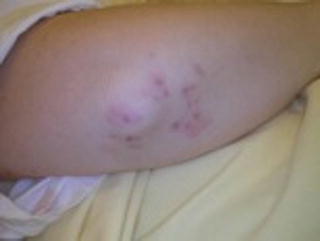
Papulonecrotic lesions are ulcerated papules that are located on the extensor surfaces of the limbs, close to elbows, knees, hands, and feet (Fig. 35.2). They can also develop on the face and scalp. Occasionally, they can resemble erythema elevatum diutinum and may be associated with IgA paraproteinemia. Present in 10 % of GPA patients, these lesions may be mistaken for rheumatoid nodules. However, unlike rheumatoid nodules, they tend to ulcerate and are mobile within the dermis [10].


Fig. 35.2
Papulonecrotic lesions on the elbow
Dermal or hypodermal nodules, the so-called subcutaneous nodules, are always palpable, typically inflammatory, tender, red, and small-sized but can sometimes be barely visible. They should be actively sought overlying lower limb vessel territories, where they can be surrounded by livedo reticularis, but are also seen elsewhere, such as the dorsal face of the upper limbs or, rarely, the trunk.
Livedo reticularis is a reddish-blue mottling of the skin in a “fishnet” reticular pattern, frequently localized on the lower limbs. It may also develop on the lower trunk and the upper limbs. It is rarely isolated in GPA and is usually associated with other cutaneous symptoms, especially nodules and necrosis.
Necrotic lesions are characterized by ulcerations or ulcers. Ulcerations of the mucosae and skin may be observed. They are more frequently nonspecific when present on mucosae [9]. Necrotic skin lesion extension and depth are highly variable, reflecting the extension and depth of histological findings, with massive skin necrosis described in a case report [11]. Extensive and painful cutaneous ulcerations may precede, by weeks to months, other systemic manifestations. Although GPA ulcers are sometimes described as “pyoderma gangrenosum-like lesions,” especially when they arise subsequent to localized trauma or the breakdown of painful nodules or pustules [6–8], they usually lack the typical raised, tender, undermined border of pyoderma gangrenosum. Sometimes numerous, they are located on the limbs, trunk, face (preauricular area), breasts (mimicking adenocarcinoma, with possible nipple retraction and galactorrhea), and perineum.
Gangrene resulting from arterial occlusion may be seen in all systemic vasculitides. It is initially characterized by the sharply demarcated blue–black color of the digits. The main differential diagnoses are thrombosis without inflammation of the vessel walls and emboli. Other concomitant cutaneous lesions can be observed in patients with skin-biopsy-proven vasculitis, e.g., nonspecific inflammation, thrombosis, and/or atheromatous embolism [7].
Clinical features that should evoke a diagnosis of cutaneous GPA in patients with pyoderma-like ulcerations include facial involvement (especially preauricular lesions), absence of typical pyoderma gangrenosum features (no undermined violaceous border, erythema, or necrotic centers), and other disease associations.
Nonspecific Dermatologic Manifestations
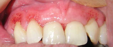
Oral ulcers are undoubtedly frequent, present in 10–50 % of GPA patients depending on the series. Unlike aphthae, they are persistent and not recurrent. Their number and localization are highly variable, having been observed on cheeks, tongue, floor of the mouth, lips, palate, gingivae, tonsils, and posterior palate (Fig. 35.3). Genital ulcers are uncommon, although penile necrosis has previously been described. Histologic findings for GPA oral ulcer tend to be nonspecific, showing only acute and chronic inflammation [9]. In a few cases, extravascular granulomas have been found [8].


Fig. 35.3
Gingival hyperplasia highly suggestive of granulomatosis with polyangiitis (Wegener’s granulomatosis)
Gingivitis. Some patients develop an unusual and distinctive gingivitis, which can suggest GPA in its early stages [12]. This gingivitis is characterized by exophytic hyperplasia with petechial flecks and a red, friable, granular appearance that begins focally in the interdental papillae and quickly spreads to produce segmental or panoral gingivitis (Fig. 35.4). It may be associated with alveolar bone loss and tooth loosening. Jaw bone pain and gingival bleeding are common. Biopsy specimens generally show nonspecific, chronic inflammation with histiocytes and eosinophils sometimes forming microabscesses. Giant cells are often present. Additional features include pseudoepitheliomatous hyperplasia with focal necrosis without vasculitis. Rarely, extravascular granuloma(s) or granulomatous vasculitis is present [8]. Differential diagnoses include drug-induced hyperplasia, or gingival hyperplasia secondary to leukemia, lymphoma, or Langerhans’ histiocytosis.
Skin necrotizing ulcerations may be nonspecific manifestations with histology finding only epitheliomatous hyperplasia, dermal hemorrhages, granulation tissue, and mixed diffuse inflammatory infiltrate(s) [6].
Erythema nodosum-like lesions consist of recurrent erythematous, edematous, and tender subcutaneous nodules. Nodule size is usually around 1 cm or 2 cm but can be much larger. Nodules are primarily located over the extensor surface of the lower limbs. They regress spontaneously without atrophic scarring. Their histomorphology in GPA is characterized by predominantly septal and focal lobular panniculitis, with a mixed inflammatory infiltrate that contains histiocytes, lymphocytes, occasional plasma cells, and singly disposed foreign-body giant cells with neutrophils. Focal lipomembranous and liposclerotic changes have also been seen without vasculitis [13].
Stay updated, free articles. Join our Telegram channel

Full access? Get Clinical Tree


