Fig. 2.1
Classification of five types of plagiocephaly according to Argenta
Type I – The cranial asymmetry is limited to the back of the skull. The degree of flattening can vary, but the deforming action is limited to this anatomical region. There is no asymmetry of the ears evaluated by measuring the nose-ear distance. The frontal bone is symmetrical, and there is no protrusion or abnormal temporal vertical stretch of the skull. This is the mildest form of positional plagiocephaly.
Type II – In this type of deformity, there are varying degrees of posterior cranial asymmetry. The deformational effect on the midline and on the basicranium is quite significant and determines the movement of the ear on the side involved in forward or down or in both directions. The asymmetry is usually more evident when one examines the baby from above. The front part of the skull is not involved and the forehead is symmetrical. There is no facial asymmetry. There are no deformity compressions of the skull. This type identifies a more severe form of positional plagiocephaly which affects not only the skull but also the rear base of the skull and the central temporal fossa.
Type III – Type III deformity includes the posterior cranial asymmetry, the malposition of the ear, and the prominence of the frontal eminence ipsilateral to the flat area. This form gives rise to a parallelogram shape of the skull classically defined as a characteristic of positional plagiocephalies and most easily noticeable by examining the child directly from above. The face is symmetrical.
Type IV – In the deformity of type IV posterior cranial asymmetry, malposition of the ear and frontal and facial ipsilateral asymmetry are present. The facial asymmetry is the result of the displacement of the adipose tissue of the cheek or, less frequently, of the hyperplasia of the ipsilateral zygomatic area. This deformity reflects the progressive nature of cranial asymmetry that involves the anterior region causing the deformation of the face.
Type V – In patients with this type of deformity, posterior cranial asymmetry, malposition of the ear, and important forehead and facial asymmetries are present. In this type, a protrusion of the temporal area and/or a vertical abnormal development of the occipital-parietal skull is also evident.
Research is required to assess the incidence of plagiocephaly using Argenta’s plagiocephaly assessment tool. A longitudinal study investigating the incidence of positional plagiocephaly at the 2-, 4-, 6-, and 12-month well-child clinic visit will provide useful information about changes in the incidence and prevalence over time, across various age ranges, and in diverse populations. The incidence of plagiocephaly in 7- to 12-week-old infants was estimated to be 46.6 % [21]. This high incidence indicates that parental education about how to prevent the development of positional plagiocephaly is warranted. The utility of using Argenta’s plagiocephaly assessment tool by public health nurses and/or family physicians needs to be established. The benefit of using Argenta’s plagiocephaly assessment tool by family physicians also needs to be ascertained. Finally, the advantages of using Argenta’s plagiocephaly assessment tool as a teaching tool for parents to track the progress of the condition after repositioning strategies are implemented need to be determined.
A classification based on pathogenetic mechanisms has been more recently proposed [22] which distinguishes PP in occipital forms (corresponding to the Argenta’s type I and II) due to compression by neurogenic hypertonicity on the bone structure of the skull and in fronto-occipital forms (Argenta’s type III, IV, and V) due to pulling forces caused by myogenic hypertonicity. This is a mere theoretical proposal that has not yet found enough applicability in clinical practice, mainly because of lack of clarity in the proposed terminology. Recent studies have shown that Argenta’s classification is a reliable and simple method for PP clinical classification. On the other hand, the minor number of categories contained in Captier’s classification would increase the reliability of the method [23].
2.2.2 Anthropometric Assessment
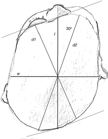
There are two main values that are used to diagnosis plagiocephaly. The first is cephalic ratio or cephalic index (CI) which is measured as the ratio of the length to width (l/w). The second is cranial vault asymmetry index (CVAI). This is the difference between the lengths of two diagonals measured 30° from the midline divided by the larger of the two diagonals. It is multiplied by 100 to create a percentage. This is commonly used because it normalizes the measurements, allowing head shapes of various sizes to be compared. This is especially important for studying PP since the head size increases greatly during the years of growth.
The classification of PP severity is also guided by quantitative assessments of skull asymmetry, expressed as transdiagonal difference for lateral PP and CI for posterior PP. Published reports of severity classification systems using these measurements vary widely, and standards remain to be established across disciplines (Fig. 2.2).


Fig. 2.2
The cranial index (CI) is the ratio l/w. The cranial vault asymmetry index (CVAI) is calculated as the difference between l1 and l2
The evaluation of the degree of PP can be performed by various methods of measurement, both direct and indirect, which allow to obtain one-dimensional, two-dimensional, or three-dimensional data.
Direct measurements are obtained using suitable calipers, according to a standardized technique useful in providing reliable results. Measurements can be performed directly on the patient or on the skull’s radiographies. This is a low-cost and easy-to-perform method. The disadvantage is due to the fact that the data obtained are exclusively of one-dimensional type, measuring distances and angles but not giving any indication as to the skull’s shape.
Accurate and consistent physical measurements aid in the diagnosis and clinical management of the infant with an abnormal skull. The value of each measurement lies in comparison with age-related norms or the individual patient at another point in time [24]. In addition to routine measurements of head circumference (occipitofrontal circumference), measurements of the infant’s cranial width, length, and transcranial diameters allow the practitioner to diagnose, classify, and monitor the presence and severity of plagiocephaly. These measurements can be taken with anthropometric spreading calipers or sliding calipers. Head length is measured at the glabella (i.e., the most prominent midpoint between the eyebrows) and the opisthocranion (i.e., the most prominent point on the occiput). These same landmarks are used to measure head circumference. Head width is measured at the maximal biparietal diameter, which may be most easily viewed from above. The infant should be upright for these measurements. The effects of thick hair can be minimized by holding the caliper points firmly against the skull [24].
Plagiocephalometri (PCM) is recently entered in clinical use: it consist in a simple and versatile, noninvading, reliable, and low-cost method, capable of quantifying the cranial deformities.
PCM measures the relationship between the transverse plane and the exact position of the ears and nose, besides the quantification of the severity of a cranial positional deformation. PCM also allows to study the time course of cranial asymmetries as well as the effects of the conservative treatment on them. PCM is performed by applying a strip of thermoplastic material around the head of the baby in the point of greatest circumference. Landmarks to the rear limit of the tragus of the ear and in the middle of the nasal bridge are then placed.
With this method, bidimensional images can be obtained, related only to the axial plane of the skull without giving any information on the parameters of its height [25, 26].
PCM provides highly reproducible and standardized anthropometric measurements in order to quantify early cranial deformities. The maintenance of a standardized position of the head is however essential to get reliable measurements. The reproducibility of anthropometric measurements is in fact essential to establish the diagnosis and the severity of the form [27].
Another method of bidimensional measurement is the photogrammetry that, although widely used, is susceptible to technical errors of measure creditable to magnification, orientation of the head, and camera-subject distance [28].
The three-dimensional photogrammetry constitutes instead an accurate, rapid, and noninvasive method that provides a promising system to get full sequential data suits on the three-dimensional aspect of the skull [29].
This is a teaching method capable of getting standardized measurements in three dimensions, combining the data of anthropometric measurements with the photographic technique.
The advantages compared to the measurements obtained with the traditional techniques (direct measurements and PCM) consist in the possibility of choice of landmarks on a static surface (without the risk of the patient moving) and in the reproducibility of measurements without causing discomfort to the patient.
The disadvantages are the cost and the size of the acquisition system: for the time being, this technique can only be performed by specialized centers. Future applications of the method are aimed at quantifying any of the volumetric changes of the skull during the follow-up [30].
Some methods using three-dimensional laser scanning, very exact but potentially harmful for the retinal function (Aldridge 2005), and methods of optic acquisition of surface have been recently employed. This technique has shorter acquisition times with a consequent decrease of the artifacts, it uses white light without risk for the retinal function, and its accuracy and reproducibility are comparable to those of PCM, of CT, and of the direct anthropometric measurements [31].
Clinical experimental studies using this method have identified four particularly important variables to assess the shape of the skull of children with PP: the index of asymmetry of the cranial vault, the index of radial symmetry, the ratio of posterior symmetry, and the ratio of total symmetry. All these variables underline an improvement obtained with the treatment, showing the utility of the optic acquisition of surface in the assessment of the treatment’s efficacy [32].
Stay updated, free articles. Join our Telegram channel

Full access? Get Clinical Tree


