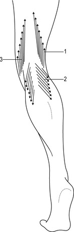
Deep fascia of the lower limbs

1.5


Deep fascia of the lower limbs


Introduction


In the literature, different fasciae such as the fascia lata, iliotibial tract, plantar fascia, crural and gluteal fasciae are described in the lower limbs but only a few words are dedicated to their macroscopic and histologic description. Recent studies highlight the unifying role of connective tissue in the limbs. In particular, these studies have demonstrated the serial continuity of the different fasciae, demonstrating how the gluteal fascia continues with the fascia lata, the crural fascia, and lastly, the plantar fascia. The deep fasciae also blend with the periosteum, tendons, and ligaments (Benjamin 2009). The many functions of the deep fascia of the lower limbs include its role as ectoskeleton for muscle attachments, and the creation of osteofascial compartments for muscles. Its role in venous return, the dissipation of tensional stress concentrated at the site of entheses, and the fact that it serves as a protective sheet for underlying structures has also been recognized.


As our understanding of the anatomy and physiology of these structures expands, the important role of the deep fasciae in the interaction among the different muscles of the limbs has become more apparent. The deep fasciae could be considered to be like a bridge, passing over the joints and the septa to connect different muscles, but recent studies (Langevin 2006a) also recognize a primary role in the perception and coordination of movements, due to the unique mechanical properties and dense innervation of these fasciae. Different researches also suggest that the deep fasciae present a basal tension. This basal tension could be due to the stretching of the underlying muscle by muscular or tendineous insertions (Stecco et al. 2009), or by the action of the myofibroblasts within the fascia (Schleip et al. 2006).


Gross anatomy


Three fundamental structures form the fasciae of the lower limbs: the superficial fascia, the deep fascia, and the epimysium.


The superficial fascia is a collagen layer that divides the hypodermis into three distinct layers: the superficial adipose tissue, a membranous intermediate layer, or true superficial fascia, and the deep adipose tissue. The thickness of the two adipose tissue layers varies in the different zones of the limbs, determining specific, regional relationships between the superficial fascia and the skin, and between the superficial and deep fasciae.


The deep fascia consists of a lamina of connective tissue that is, generally, easily separable from both the underlying muscles and the overlying superficial fascia. In fact, there is virtually an uninterrupted plane of gliding between the deep fascia and the muscles surrounded by their epimysium, with just a little layer of interposing, loose connective tissue to facilitate gliding. This loose connective tissue appears as a pliable, gel-like gelatinous substance. Histologic studies demonstrate that fibroblasts are widely dispersed within this tissue and that collagen and elastic fibers are disposed in an irregular mesh.


A few strong intermuscular septa originate from the inner surface of the deep fascia of the lower limbs and extend between the muscle bellies, dividing the thigh into different compartments, and providing an origin to some lower limb muscle fibers (Plate 1.5.1).


It is important to recognize that even though the deep fascia of the lower limbs in the thigh is called fascia lata, and in the leg crural fascia, it is actually the same structure. This fascia appears as a thick, whitish layer of connective tissue, similar to an aponeurosis, with an average thickness of 1 mm and, in general, the deep fascia of the lower limbs is thicker in the posterior regions of the limbs. Nevertheless, studies concerning the variations in thickness of the lower limb fasciae have demonstrated some regional differences. In particular, the deep fascia of the anterior thigh presents a mean thickness of 944  ±  102 μm. It is thinner in the proximal region (541  ±  23 μm) and thicker near the knee (1419  ±  105 μm), while in the middle third of the thigh it presents a mean thickness of 874  ±  62 μm. In the lateral region, it is reinforced by the iliotibial tract. The iliotibial tract is not separable from the deep fascia by dissection. Therefore, anatomically, it can not be considered a separate entity, but a reinforcement of the lateral aspect of the fascia lata.


The crural fascia has an average thickness of 880 μm, which progressively decreases from 1 mm in the popliteal region to 700 μm in the distal third of the leg.


Around the knee and the ankle, the deep fascia is reinforced by additional fibrous bundles, commonly called retinacula; however, it should be emphasized that in all of the fasciae many fibrous bundles running in different directions are macroscopically visible.


The retinacula


The retinacula are typically regional specializations of the deep fascia; in particular, they are thickenings of the deep fasciae and, as such, are not separable. They appear as a strong fibrous bundle with a mean thickness of 1372 μm and a criss-cross arrangement of the collagen fibers. The retinacula have many bone insertions, and these entheses may be fibrocartilaginous. At other points, they can glide over the bones thanks to interposing loose connective tissue between the retinacula and the periosteum. From a functional point of view, the retinacula of the ankle have classically been considered as a pulley system, maintaining the tendons adherent to the underlying bones during movements of the tibiotarsal joint, and as important elements for ankle stability, connecting various bones. However, in 1984, Viladot et al. stated that they may play an important role in proprioception. For example, the peroneal retinacula may be stretched by inversion of the ankle joint, activating reflex contraction of the peroneal muscles. Ankle retinacula can be easily evaluated by magnetic resonance imagery, as they appear as low signal intensity bands, sharply defined in the context of the subcutaneous tissue in T1-weighted sequences, with a mean thickness of 1.25 mm (SD  ±  0.198). Retinacula can also be subjected to traumatic ruptures, sometimes resulting in subluxation of the underlying tendons. Also the knee retinacula could be easy evaluated by magnetic resonance imaging, as they appear clearly as low signal int-ensity bands. In patients affected by patellar femoral malalignment or anterior knee pain, differences in thickness, innervations, and vascularisation are appreciable.


Fibrous expansions and muscular insertions


In a few specific regions, the muscles of the lower limb connect with the deep fascia via fibrous expansions or direct insertions of their muscular fibers. These expansions and insertions are well worth some in-depth discussion for their potential functional implications.


While the iliotibial tract could be considered the tendon of the tensor fascia lata and the gluteus maximus muscles, it is also a reinforcement of the fascia lata. It has extensive attachments to the lateral intermuscular septum in the thigh and many muscular fibers of the vastus lateralis muscle also originate from this septum. Therefore, during movement of the lower limb, the lateral intermuscular septum is stretched proximally by the gluteus maximus, and distally by the vastus lateralis muscles (Plate 1.5.2). Distally, the iliotibial tract is attached to Gerdy’s tubercle at the upper end of the tibia, but it also has an expansion into the antero-lateral portion of the crural fascia. Fairclough et al. (2007) suggest that iliotibial band syndrome is not due to frictional forces created by moving forwards and backwards over the tibial condyle during flexion and extension of the knee, but to tensional changes within the iliotibial tract itself. Similarly, the sartorius, gracilis, and semitendinosus muscles form the pes anserinus in the medial portion of the knee, but they also extend some expansions into the medial aspect of the crural fascia (Fig. 1.5.1). Besides, the quadriceps muscle has some obliquely directed fascial expansions arising from the vastus medialis and lateralis muscles, that pass anterior to the patella fusing with the fascia lata, and contributing to knee retinacula formation, and the distal tendon of the semimembranosus muscle has two expansions in the popliteal region: one extends into the posterior wall of the knee joint capsule, forming the oblique popliteal ligament, and one extends into the fascia of the popliteus muscle. Distally, the proximal portions of the gastrocnemius muscle are inserted directly onto the fascia, so that these muscular fibers could be considered as tensors of the fascia (Plate 1.5.3), and anteriorly the tibialis anterior muscle and flexor hallucis longus insert onto the overlying fascia and the intermuscular septum. In this way, around the knee, it is quite difficult to separate the deep fascia from the underlying muscles and tendons.


Stay updated, free articles. Join our Telegram channel

Aug 24, 2016 | Posted by in ORTHOPEDIC | Comments Off on Deep fascia of the lower limbs

Full access? Get Clinical Tree

Get Clinical Tree app for offline access