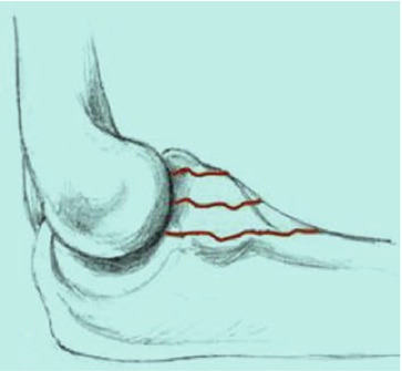
Type I
Fracture of the tip
Type II
Fracture involves less than 50 %
Type III
Fracture involves more than 50 % of the coronoid
Each type is subclassified according to the absence (A) or presence (B) of a dislocation (Fig. 9.1).


Fig. 9.1
Regan and Morrey classification
In 2003, O’Driscoll et al. [1] introduced a new classification system with special consideration of the anteromedial facet (Table 9.1). Type 1 fractures affect the tip, type 2 the anteromedial facet, and type 3 the base. Each type is divided into subtypes. Treatment options can be derived from this classification.
Table 9.1
Classification of coronoid fractures developed by O’Driscoll et al.
Type | Fracture | Subtype | Description |
|---|---|---|---|
I | Tip | 1 | ≤2 mm of the tip |
2 | >2 mm of the tip | ||
II | Anteromedial facet | 1 | Anteromedial rim |
2 | Anteromedial rim + tip | ||
3 | Anteromedial rim + sublime tubercle + tip | ||
III | Basal | 1 | Olecranon basal coronoid fractures |
2 | Transolecranon basal coronoid fractures |
9.4 Treatment
Most coronoid fractures are small type I fractures of the tip. These can be treated conservatively. The treatment of larger coronoid fragments is usually operative, although the fracture may seem harmless on standard radiographs. As these fractures must be interpreted as fracture-dislocations in most cases, even fractures that appear harmless on standard radiographs may present severe injuries. Correct classification requires a CT scan. Not only the bony structures, but also the soft tissues have to be addressed during surgery.
Stay updated, free articles. Join our Telegram channel

Full access? Get Clinical Tree


