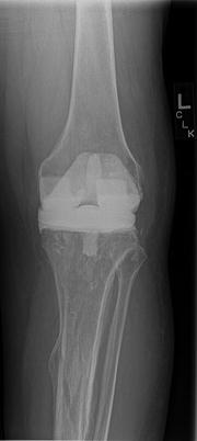
Fig. 4.1.
(a and b) AP radiographs reveal posttraumatic arthritis with retained lateral tibia locking plate with broken distal cortical screws.
4.1.1 Diagnosis
The patient presented with severe posttraumatic arthritis and retained hardware. Despite a benign history and knee aspiration, the serologic evaluation was concerning for occult infection. The knee aspiration, although negative, may be unreliable due to the extra-articular nature of the hardware. Consideration was made to perform a staged approach to the reconstruction with removal of hardware and antibiotic treatment prior to definite reconstruction in anticipation of periprosthetic infection.
4.1.2 Management
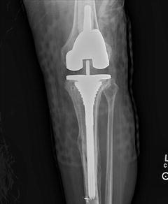
The patient underwent a resection arthroplasty and complete removal of the hardware with placement of an articulating antibiotic cement spacer (see Fig. 4.2). Intraoperative frozen sections of the synovial tissue and perihardware tissue revealed significant acute inflammation with microabscesses consistent with infection. A universal screw removal set and high-speed metal-cutting burrs were required for complete hardware removal. Intraoperative cultures were negative and the patient received 6 weeks of broad-spectrum intravenous antibiotics, after which his serologic markers normalized. Final reconstruction was performed thereafter using stemmed implants to bypass stress risers (see Fig. 4.3).


Fig. 4.2.
Postoperative AP radiograph with complete hardware removal and resection arthroplasty with articulating antibiotic cement spacer.

Fig. 4.3.
Postoperative AP radiograph after arthroplasty reconstruction with long-stem components.
4.1.3 Literature Review
4.2 Preoperative Considerations
Complete evaluation of the posttraumatic knee requires a through history of the events surrounding the prior injury, surgery, and any perioperative complications. The physical examination includes evaluation of prior incisions and baseline range of motion, ligamentous stability, and skeletal deformity. Radiographic evaluation consists of a standard weight-bearing knee series as well as full-length femur and tibia views, if necessary, to visualize hardware completely as well as any extra-articular deformities.
4.2.1 Periprosthetic Infection
Periprosthetic joint infection remains a feared complication with significant negative implications for the patient and surgeon alike. Although the typical incidence of periprosthetic infection is quoted as <2 % in the primary setting, this number can exceed 12 % in high risk groups [2]. Suzuki et al. reported that previous open reduction with internal fixation and retained internal fixation hardware was found to be an independent risk factor to the development of periprosthetic joint infections. In contrast to this study, however, Klatte et al. found no increased risk of periprosthetic joint infection in patients with pre-existing osteosynthesis hardware at 5.4 years follow-up of 115 patients they studied [3]. Regardless, the debilitating consequences of periprosthetic joint infections require one to maintain a high index of suspicion of occult infection in the presence of previous internal fixation. History of open fractures, persistent wound drainage, and previous antibiotic treatment following ORIF should raise concerns of chronic infection. Routine serologic evaluation with a complete blood count, erythrocyte sedimentation rate, and C-reactive protein should be obtained. Elevated values and/or high clinical suspicion should prompt a knee aspiration. It is important to recognize, however, that a negative aspiration does not eliminate the possibility of an infection of in situ hardware, as the hardware may be extra-articular. Additional nuclear medicine imaging, such as a white blood cell labeled scan, can be of value if the diagnosis is in question. A positive infection workup, or high clinical suspicion, should trigger a two-stage approach with complete hardware removal, thorough debridement, and tailored intravenous antibiotic treatment. The arthroplasty procedure should be delayed until infection has been ruled out or resolved.
4.3 Surgical Planning
4.3.1 Hardware Management
As a general rule, we recommend removing symptomatic hardware or that which interferes with the preparation for, or implantation of, the total knee arthroplasty. Complete removal is often unnecessary in many instances. In general, this can be accomplished in a single-stage operation. This allows for less surgical morbidity, decreased soft tissue dissection, and prevents creation of unnecessary stress risers. When multiple incisions are anticipated for hardware removal, a staged approach should be entertained to allow the soft tissues to heal prior to the arthroplasty procedure. In order to facilitate hardware removal, complete understanding of implants in place, their location, and any posttraumatic deformities should be part of the surgical planning. To do so, full-length radiographic evaluation of the tibia and femur is often required. In addition, previous operative reports can clarify what specific implants are in place, so that appropriate removal sets can be requested. Often, however, the hardware is unidentified or damaged and one must therefore be equipped with comprehensive hardware extraction sets. A universal screw removal set and high-speed cutting burrs (e.g., Midas Rex) can facilitate hardware extraction and should be available (see Fig. 4.1b). The presence of hardware does not simply provide an obstacle in its removal, but also in implant selection for the arthroplasty.
Stay updated, free articles. Join our Telegram channel

Full access? Get Clinical Tree


