Fig. 5.1.
Preoperative long-standing film showing preoperative plan.
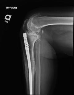
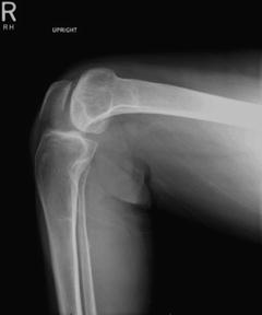
Fig. 5.2.
Preoperative lateral film.
5.1.2 Management
Patient underwent left lower extremity hardware removal and total knee arthroplasty in a single surgery. The varus deformity was significant and the superficial MCL was released in order to balance the knee. A larger than normal polyethylene insert was needed to fill the flexion and extension gaps after the aggressive medial release. We were prepared to increase the constraint, but this was not necessary. The deformity was corrected intra-articularly without the use of computer navigation or the requirement for extra-articular correction.
5.1.3 Outcome
The patient recovered uneventfully following our standard TKA postoperative protocol. She is doing well with a ligamentously stable knee and a postoperative range of motion of 0–105° (Figs. 5.3 and 5.4).
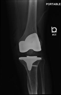
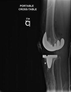
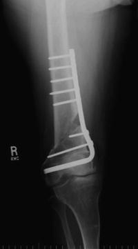
Fig. 5.3.
Postoperative AP film showing intra-articular correction.
Fig. 5.4.
Postoperative lateral film.
5.2 Literature Review
Lower extremity extra-articular deformity can occur due to prior trauma, surgery, metabolic bone disease, or congenital malformations. It can be complicated by many things, such as previously placed hardware and prior incisions. These interfere with instrumentation and alter landmarks. Long-standing deformity leads to soft tissue contractures requiring releases. The goals of performing a TKA in a person with extra-articular deformity are the same as those for a TKA with the diagnosis of idiopathic osteoarthritis. One needs to establish a neutral mechanical axis and a ligamentously balanced and stable knee throughout a functional arc of motion. In general, deformities of the femur are more difficult to deal with than deformities of the tibia due to the intramedullary instrumentation of the femur used with conventional instrumentation.
When faced with these challenging cases, there are a few basic principles that need to be addressed. Most importantly the patient needs to be counseled on the increased risk of complications and decreased functional outcomes of these surgeries compared to primary cases. Full-length extremity films need to be obtained to preoperatively plan for deformity correction and a CT may be considered to evaluate rotational deformity. Indolent infection should be ruled out with screening labs (ESR and C-reactive protein) and possible joint aspiration if serologies are elevated or there is a high clinical suspicion for infection. Hardware removal does not need to be staged with the exception of a diagnosed infection and only the hardware that interferes with surgery should be removed. If possible, previous incisions should be used and old incisions should be crossed at right angles.
The first decision that needs to be made is if the deformity is correctable with intra-articular bony cuts and ligamentous releases or is an extra-articular correction required. The amount of correction needed is positively correlated to the severity of the deformity and inversely correlated to the distance of the deformity from the joint line [1]. A small deformity of the proximal femur is more easily corrected than a large deformity of the distal femur. Intra-articular correction is the most desirable scenario because it is more familiar to the surgeon and more efficient. A previous study has determined that intra-articular correction is possible if the bony resection is <20° in the femoral coronal plane, <25° in the femoral sagittal plane, and <30° in the tibial coronal plane [2]. Most importantly however, these resections should not compromise the insertions of the collateral ligaments. Intra-articular correction also eliminates problems associated with an osteotomy, which are discussed later. The decision of intra vs. extra-articular correction is based on the amount of correction desired and determined on the full-length standing hip-knee-ankle film. If the amount of bony resection will compromise the insertions of the collateral ligaments, then an extra-articular osteotomy is necessary.
Extra-articular osteotomies allow a larger correction but bring about the possibilities of osteotomy nonunion and decreased weight-bearing status that may interfere with rehabilitation after TKA. Osteotomies may also require additional incisions in an already poor soft tissue envelope. The traditional operative procedure is a two-stage approach of osteotomy and fixation followed by TKA after the osteotomy has healed (Figs. 5.5, 5.6, 5.7, 5.8, 5.9, and 5.10). Some surgeons have attempted a one-stage surgery with intraoperative computer navigation with mixed results [3–5]. Older studies warn that simultaneous surgery produces inferior outcomes [6, 7]. Most of these surgeries were performed without computer navigation assistance or stemmed implants. One computer-assisted study evaluating a subset of procedures that required a simultaneous osteotomy (3/40 TKAs) showed no outcome differences or increase in complications [3]. Worse outcomes and increased complications have been shown when simultaneous surgery is performed without navigation [4].
 Periprosthetic Infection: Management of Late Acute Hematogenous Infection
Complex Primary Total Knee Arthroplasty: Management of Previous Hardware (Posttraumatic OA)
Revision Total Knee Arthroplasty: Management of Deficient Extensor Mechanism
Complex Primary Total Knee Arthroplasty: Management of Varus Knee
Periprosthetic Infection: Management of Chronically Infected Total Knee Arthroplasty
Complex Primary Total Knee Arthroplasty: Management of Flexion Contracture
Periprosthetic Infection: Management of Late Acute Hematogenous Infection
Complex Primary Total Knee Arthroplasty: Management of Previous Hardware (Posttraumatic OA)
Revision Total Knee Arthroplasty: Management of Deficient Extensor Mechanism
Complex Primary Total Knee Arthroplasty: Management of Varus Knee
Periprosthetic Infection: Management of Chronically Infected Total Knee Arthroplasty
Complex Primary Total Knee Arthroplasty: Management of Flexion Contracture


Fig. 5.5.
Preoperative AP film showing significant coronal plane deformity of both femur and tibia.
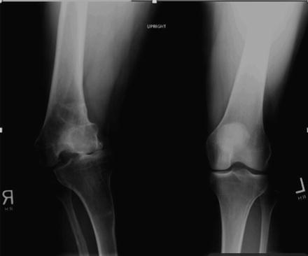
Fig. 5.6.
Preoperative lateral film showing minimal sagittal plane deformity.
Related posts:
Periprosthetic Infection: Management of Late Acute Hematogenous Infection
Complex Primary Total Knee Arthroplasty: Management of Previous Hardware (Posttraumatic OA)
Revision Total Knee Arthroplasty: Management of Deficient Extensor Mechanism
Complex Primary Total Knee Arthroplasty: Management of Varus Knee
Periprosthetic Infection: Management of Chronically Infected Total Knee Arthroplasty
Complex Primary Total Knee Arthroplasty: Management of Flexion Contracture
Stay updated, free articles. Join our Telegram channel
Full access? Get Clinical Tree
