CHAPTER 29 Complex Instability of the Elbow
INTRODUCTION
Since the first edition of this book, improvements in the recognition and treatment of complex instability are among the most significant developments in managing elbow trauma. The coexistence of multiple articular and soft tissue injuries is being increasingly appreciated.9,42
DEFINITION
Complex instability of the elbow is defined as an injury that destabilizes the elbow because of damage to the articular surface and to the ligamentous structures.21 Others add soft tissue involvement, including neural and vascular injuries in the diagnosis.36 However, vascular injuries are uncommon in this setting. Over a 10-year period, for example, Moneim and Garst20 documented only two instances of vascular injury among 56 with fracture dislocation injury. The clinical presentation is usually one in which the instability is obvious, but more subtle forms are being recognized.39 In this chapter, the relative contributions of the articulation and the ligaments to normal stability, as well as to their interactions, are first defined. We then offer a rationale for the reliable treatment of a spectrum of these acute injuries.
CONTRIBUTIONS TO NORMAL STABILITY
ARTICULAR ELEMENTS
Although the distal humerus is sometimes involved, for the purpose of this discussion, it is assumed that the distal humerus is intact. Therefore, the articular elements to be considered are the radial head, olecranon, and coronoid. The relative role of these elements to elbow stability have been recently reviewed.27
Radial Head
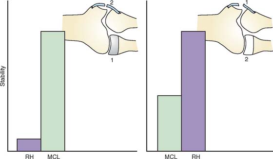
It is known clinically that the radial head may be resected without altering the stability of the otherwise normal elbow.27 Therefore, the contribution of the radiohumeral joint to elbow stability is intimately related to and dependent on the integrity of the collateral ligaments. Experimental data have clearly documented that the resistance to valgus stress provided by the radial head is minimal when the medial collateral ligament is intact24,25,40 (Fig. 29-1). However, the radiohumeral joint offers enough resistance to valgus stress to prevent subluxation if the medial collateral ligament is attenuated or torn. The major structure resisting initial valgus displacement, even with an intact radial head, is the medial collateral ligament. If the medial collateral ligament is intact, the radial head offers little resistance to valgus stress, but if the medial collateral ligament is attenuated or torn, the radial head assumes the role of an important stabilizer. Thus, the radial head is an important secondary stabilizer to valgus stability.
The relationship of the radial head to the ulnar part of the lateral collateral ligament has been initially defined experimentally by O’Driscoll and associates.30,32 Today, posterolateral rotatory instability is well recognized to be associated with lateral ulnar collateral deficiency5,19,33 and can occur in the presence or absence of the radial head. However, clinical experience suggests that elbows without a radial head do less well after reconstruction of the ulnar part of the lateral collateral ligament than do those in which the radial head is intact.2,29 This provides further evidence of the important role of the radial head to resist posterolateral rotatory instability but, once again, in a secondary capacity.
Olecranon
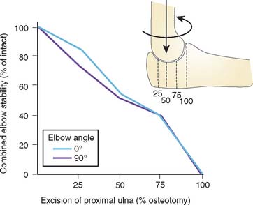
The major determinant of stability of the elbow is clearly the ulnohumeral joint, specifically the coronoid. Although the stabilizing influence of this joint has not been studied to any great extent, the relative contribution of the olecranon in resisting various loading configurations has been shown to be linearly correlated with the extent of resection of the proximal part of the ulna (Fig. 29-2).1 The critical amount of articulation required for maintaining stability is about 50%. These data may be altered by dynamic forces, but this has not been studied.
Coronoid
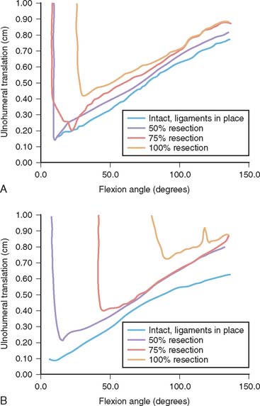
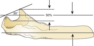
The amount of the coronoid required for stability with or without ligamentous integrity and with and without the radial head is just now emerging. Experimental and clinical experience suggests that at least 50% of the coronoid must be present for the ulnohumeral joint to be functional (Fig. 29-3). Absence of the radial head further and dramatically compromises the elbow with a 50% coronoid deficiency. From a practical perspective, we have found it useful to observe that the typical 30-degree angle formed by a line from the intact olecranon and coronoid is reduced to 0 degrees when the critical50% of the coronoid is present (Fig. 29-4). Anatomically and clinically, the coronoid is the subject of intense investigation to better understand fracture patterns, and the “critical portion” needed for stability.4
LIGAMENTOUS CONTRIBUTIONS
The relative contributions of the medial and lateral collateral ligaments to varus-valgus stability with the elbow articulation intact and in flexion and extension have been studied experimentally.23 This investigation has shown that the collateral ligaments provide approximately 50% of the varus-valgus stability of the joint, and the articular surfaces provide an additional 50%. The only exception is with the elbow in full extension: under this condition, the ulnohumeral joint and anterior capsule render the elbow stable to varus-valgus stress, even in the absence of collateral ligaments. The role of the ligaments in conjunction with the articulation is described earlier.
CLINICAL MANAGEMENT: PRINCIPLE
A basic principle underlying the treatment of complex instability is simply that a competent ulnohumeral jointbe attained and maintained.21,31 This is the primary overriding principle.
FRACTURE OF THE RADIAL HEAD WITH ATTENUATION OR TEAR OF THE MEDIAL COLLATERAL LIGAMENT
The presentation of medial ligament injury can be extremely subtle. The prevalence is difficult to ascertain, but in my experience, as an isolated lesion, it has occurred only in about 1% to 2% of patients who had a fracture of the radial head.22 One should have a highlevel of suspicion that an injury to the ligament might be present when there is a compression fracture with or without comminution of the radial neck. Ecchymosis on the medial side of the elbow should be an obvious clue but is often absent.
Treatment
Treatment of this difficult structured problem is based on the realization that the medial collateral ligament tends to heal if the ulnohumeral joint is reduced. Furthermore, the radial head is an important secondary stabilizer to valgus stress when the medial collateral ligament has been disrupted. The primary goal of the treatment strategy is to restore ulnohumeral joint stability. However, a reconstructed medial collateral ligament has been observed to stretch in the absence of radial humeral integrity; hence, if it is amenable to fixation, the fracture is fixed; if not, the head is replaced. If the arc is stable within 40 degrees of extension, unrestricted passive motion is allowed after 7 to 10 days of protection. If dislocation occurs with extension of approximately 60 degrees, the elbow is immobilized for 10 days, after which motion in a Mayo Elbow Brace with a 45-degree extension stop, is allowed for 2 weeks. In our practice, the collateral ligament need not be repaired in those with a stable ulnohumeral, radiohumeral joint. Nonetheless, the popularity of suture anchors has prompted a more aggressive attitude toward ligament repair. Rodgers and colleagues38 report 88% satisfactory results in 17 patients treated with suture anchors who had major elbow injuries.
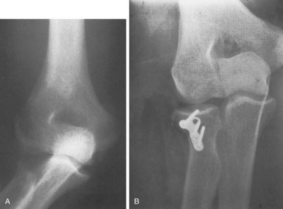
If osteosynthesis is not possible, then restoration of the radiohumeral joint with the use of a prosthesis becomes a higher priority. Prostheses of today are considerably more effective than in the past. Therefore, the possibility of use of metal implants has been reintroduced.7,12,15 In some instances in which the medial collateral ligament has been disrupted, replacement of the radial head with an allograft has been performed as an alternative method of providing stabilization (Fig. 29-5A and B).
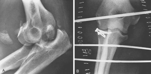
Finally, a hinged external fixator is applied in those circumstances in which it is desirable to protect a fractured radial head, the coronoid, or to protect a ligament repair (see Chapter 33).3,13,17 There are several articulated fixators available, but for this indication, Kamineni has shown that the simple half-pin configuration of the Dynamic Joint Distractor (DJDII) (Stryker, Mahwah, NJ) is effective to stabilize the joint even in the absence of both collateral ligaments. Hence, because of cost and simplicity, this is our choice of devices (Fig. 29-6).13 If the fixator is not used, patients are fitted with the Mayo Brace in the locked mode for protection for about 2 weeks postoperatively; the brace is then unlocked, and motion is allowed in the brace through the stable arc. The hinged brace is worn for a total of at least 6 weeks or until the joint is considered stable.
Stay updated, free articles. Join our Telegram channel

Full access? Get Clinical Tree


